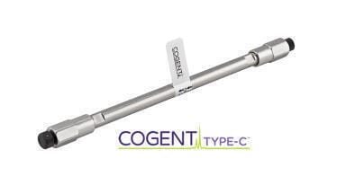
Columns (LC)
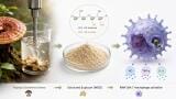
Researchers in China have developed a high-throughput chromatographic strategy that can identify more than 2,300 proteins from a single hepatocyte slice in five minutes, offering a faster route to single-cell spatial proteomics and tumour microenvironment analysis
A research team in China has introduced a high-throughput strategy for single-cell spatial proteomics that uses an ordered colloidal crystal chromatographic column to accelerate analysis while preserving deep proteome coverage.
The study was led by Professor Zhang Lihua and Professor Liang Yu from the Dalian Institute of Chemical Physics of the Chinese Academy of Sciences in the city of Dalian, Liaoning, China, with the work representing a significant technical advance for spatial proteomics at single-cell resolution, a field that seeks to map how proteins are distributed across biological tissues and how this molecular organisation relates to health and disease.
Spatial proteomics has become an important approach to understand biological function because proteins are not only defined by their abundance but also by their location. The position of proteins within and between cells can influence signalling pathways, tissue organisation and disease processes. At single-cell resolution, this type of analysis is especially valuable because it can reveal cellular heterogeneity, identify differences between neighbouring cells and help researchers to investigate interactions within complex tissue microenvironments.
However, current approaches face a major practical limitation. Mainstream spatial proteomics often combines nano-liquid chromatography–mass spectrometry with laser capture microdissection. Laser capture microdissection allows researchers to isolate defined regions or individual cells from tissue sections, while nano-liquid chromatography–mass spectrometry separates and identifies proteins with high analytical sensitivity. Yet as spatial resolution increases, the number of tissue sections or microscopic sampling points also rises sharply. This creates a severe throughput bottleneck, particularly for large-scale tissue studies that require the analysis of many samples.
Even with advanced chromatographic columns and high-performance mass spectrometry systems, single-cell proteomic analyses typically require about 30 minutes for each sample to achieve sufficient proteome coverage. That analysis time can become prohibitive when researchers need to build molecular atlases or compare multiple tissue regions across disease stages.
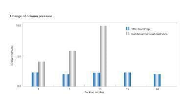
To address this challenge, the research team developed a novel colloidal crystal chromatographic column based on the ordered assembly of 800 nm monodisperse C18 colloidal particles. C18 refers to an octadecylsilane surface chemistry widely used in reversed-phase liquid chromatography to separate peptides and proteins according to hydrophobicity. The researchers designed the column to exploit a highly ordered architecture, submicrometre particle size and non-porous structure. Together, these features allowed the column to achieve an efficiency of more than two million ‘theoretical’ plates per metre, about 10 times higher than that of conventional sub-2 μm chromatographic columns.
Column efficiency – expressed as theoretical plates per metre – reflects how effectively a chromatographic column can separate analytes. Higher efficiency generally means sharper peaks, better resolution and greater analytical performance, which is particularly important when sample amounts are extremely limited, as in single-cell proteomics.
When the team applied the method to single-cell-resolution spatial proteomics, it achieved rapid and deep protein identification. The approach enabled researchers to identify up to 2,304 proteins from a single hepatocyte slice within five minutes. Even under a two-minute gradient, the method still identified more than 1,000 proteins. This performance suggests that the ordered colloidal crystal column could substantially increase throughput without sacrificing the proteome depth required for meaningful biological interpretation.
The researchers then applied the method to hepatocellular carcinoma tissue which is the most common form of primary liver cancer and is marked by pronounced biological heterogeneity, both within tumours and between tumour regions. Using the novel chromatographic approach, the team rapidly characterised the spatial proteomic features of liver regions, early-stage hepatocellular carcinoma regions and advanced tumour regions.
The analysis also revealed proteomic heterogeneity among distinct cell populations within tumour regions at single-cell resolution. Such heterogeneity can be biologically and clinically important because different tumour cell populations may vary in their metabolism, signalling activity, immune interactions or treatment response. A faster method to profile these differences could therefore support more detailed molecular atlas construction and improve the study of disease tissue microenvironments.
“Our study provides a high-throughput technological tool for single-cell spatial proteomics, molecular atlas construction, and investigations of disease tissue microenvironments,” Zhang concluded.
For further reading please visit: 10.1002/anie.8045649