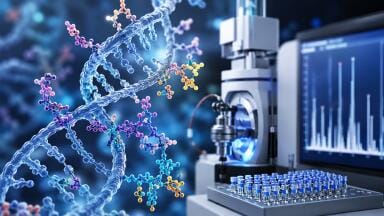
![Figure 2: Analysis of empty LNPs spiked with mRNA at two different pH conditions: pH 4 and pH 7.5 [1].](/assets/file_store/pr_files/66042/thumbnails/images/768w-432h-t-fit-251015_ebulletin_LNPs_Fig2.jpg)
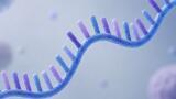
![Figure 3: Comparison of two samples where the EE outcomes align (blue) and where both methods diverge significantly (orange) [1].](/assets/file_store/pr_files/66042/thumbnails/images/768w-432h-t-fit-251015_ebulletin_LNPs_Fig3.jpg)
Liquid chromatography
To enhance intracellular delivery and preserve mRNA integrity against RNase-mediated degradation, therapeutic mRNA can be encapsulated in lipid nanoparticles (LNPs). These LNPs transport the payload directly into the cytoplasm, enabling efficient translation. The encapsulation efficiency (EE) defines the proportion of mRNA that will eventually be delivered into the cells and is therefore directly linked to the therapeutic potential of the drug product. Current workflows often rely on spectrofluorimetric detection using fluorescent dyes such as RiboGreen (Thermo Fisher Scientific), which bind to mRNA. However, this method has several drawbacks such as it is prone to matrix effects, the binding of the dye to mRNA is highly sensitive to environmental factors or the structure of the LNP can be affected by dilution. As LNPs exhibit neutral charge, anion exchange chromatography (AEX) offers a targeted strategy to separate free mRNA from unretained drug product.
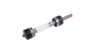
This Technical Note based on the publication and data of the University of Geneva and Sanofi’s mRNA Center of Excellence in France demonstrates that the AEX method using a bioinert coated YMC Accura BioPro IEX QF column provides a refined and more robust alternative to commonly applied analytical workflows [1].
In some drug product (DP) samples a second pre-mRNA peak was observed which absorbed across all UV-wavelengths. The absorbance ratio of 260/230 nm confirms the assumption of an mRNA–LNP within the analysed sample. This phenomenon does not appear when the empty LNP or the empty together with a DS spike is injected. But it appears when injecting the empty LNP spiked with DS at an acidic pH (Figure 2). The data suggest that mRNA binds to the positively charged surface of the LNP. To improve the separation of the free mRNA and the pre-mRNA peak an isocratic step was implemented. Light-scattering effects currently prevent the quantification of the pre-mRNA peak using standard detection methods. However, structural information about the LNP can still be derived on a qualitative level [1].
The comparison of both analytical approaches reveals consistent results in many cases, while the RiboGreen method occasionally reports a notably lower EE. Figure 3 displays the chromatograms of two selected samples where the outcomes align (blue) and where both methods diverge significantly (orange). The blue sample produces a peak exclusively for free mRNA, while the orange sample additionally reveals a pre-mRNA signal. The data suggest that the RiboGreen dye binds to surface-associated mRNA, which falsely contributes to the free mRNA signal and results in a reduced EE.
The AEX method accelerates analysis, reduces cost, and yields additional structural insights into the LNP, positioning it as a viable alternative for determining EE. The study provides evidence for the presence of surface-associated or transmembrane mRNA within specific formulations. Further research must establish whether this mRNA fraction retains therapeutic functionality.
Find more details in the Technical Note.
[1] Athanasios Tsalmpouris, Sofiane Mahjoubi, Camille Malburet, Chamsan Daher-Hassan, Marc François-Heude, Jean-François Cotte, Davy Guillarme, and Jonathan Maurer Analytical Chemistry 2025 97 (35), 19275-19282; DOI: 10.1021/acs.analchem.5c03299