HPLC, UHPLC
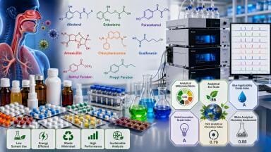
Researchers have mapped the chemical profile of Viola odorata (sweet violet) flowers using advanced chromatography and mass spectrometry. The study identified flavonoids and anthocyanins with strong anti-inflammatory and cytotoxic effects in human cell models, including potent inhibition of COX and 5-LOX enzymes. By isolating pure compounds such as kaempferol 3-O-rutinoside, the work provides evidence that sweet violet could form the basis of future plant-derived anti-inflammatory or anticancer therapies
A chromatography-led investigation has mapped the polyphenolic chemistry of Viola odorata (sweet violet) flowers and has linked discrete components to anti-inflammatory and cytotoxic effects in human cell models. Sweet violet has a long history in traditional medicines – from Ancient Greece to the Indian Ayurveda via Persia and the Arabic world – and used for treating respiratory conditions, inflammation and skin disorders.
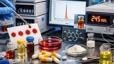
The study placed separation science at the centre of its workflow: high performance liquid chromatography (HPLC) and ultra-performance liquid chromatography/electrospray ionisation–mass spectrometry (UPLC/ESI–MS) defined the chemical space, while preparative steps and thin-layer confirmation delivered purified flavonoids for bioassay and computation.
The authors set out to close a gap in the literature by producing a flower-specific chemical atlas and by tying that atlas to function. They first quantified total phenolics, flavonoids and anthocyanins in a methanolic extract, then used chromatographic resolution to assign identities and amounts.
The extract contained 81.34 ± 0.17 mg gallic acid equivalents per gram of extract for total phenolics, 69.45 ± 0.24 mg catechin equivalents per gram for total flavonoids, and 92.43 mg cyanidin-3-glucoside per 100 g for anthocyanins. Those bulk values established a high-polyphenol baseline and justified deeper separation.
Analytical HPLC on a C18 platform resolved twelve phenolic acids and ten flavonoids with diode-array detection across 280, 320 and 360 nm. Identification rested on retention-time matches to authenticated standards and corroborating ultraviolet spectra. Gentisic acid was present in the highest amount among phenolic acids at 391.37 μg/g. Among flavonoids, apigenin-7-glucoside (417.22 μg/g), catechin (372.56 μg/g) and rutin (262.73 μg/g) dominated. Gradient design and a moderate 0.8 ml min⁻¹ flow balanced peak capacity with analysis time and produced baseline separation for the abundant constituents.
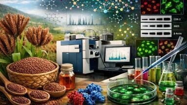
To widen coverage and assign structures beyond what photodiode arrays could support, the team chose to use UPLC/ESI–MS in both positive and negative modes. Short sub-2 μm C18 media, a binary formic acid system – and controlled ramps in organic content cut run time and sharpened band shapes – which improved signal-to-noise for low-abundance species.
Exact-mass data and characteristic fragmentation pathways then established identifications. In total, the UPLC/ESI–MS catalogue comprised eight phenolic acids and derivatives, three flavonols (including quercetin, myricetin and isorhamnetin), four flavones (including luteolin and methoxyflavones), fourteen flavonoid glycosides, and five anthocyanins derived from cyanidin, delphinidin and petunidin. Violanin – delphinidin-3-(4″-p-coumaroyl)-rutinoside-5-glucoside – served as a marker anthocyanin, with a molecular ion at m/z 920 in positive mode and a diagnostic aglycone fragment at m/z 303.
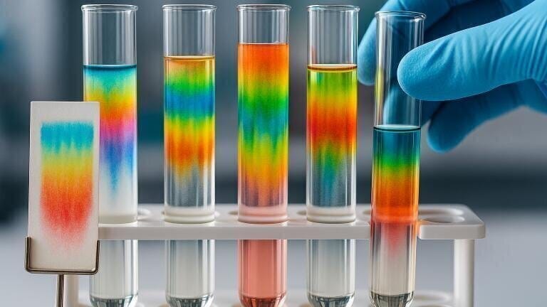
A silica column with stepwise polarity and confirmatory thin-layer chromatography yielded three reference-grade flavonoids from the complex matrix: 5,7-dihydroxy-3,6-dimethoxyflavone, luteolin 7-O-glucoside, and kaempferol 3-O-rutinoside. Ultraviolet bathochromic shifts with complexing reagents, consistent melting points, and complete proton and carbon nuclear magnetic resonance spectra verified each structure against the literature. By firmly basing biological testing to chromatographically pure substances, the study reduced attribution errors that can plague extract-only reports.
With the chemical observational data available, the investigators assessed activity at pro-inflammatory enzyme targets and across three human cell lines. Enzyme assays used commercial cyclooxygenase 1 and 2 (COX-1 and COX-2) kits of ovine/human origin and a human recombinant 5-lipoxygenase (5-LOX) kit, with indomethacin and zileuton as internal controls. The flower extract inhibited all three enzymes with half-maximal inhibitory concentration values near the benchmarks.
Among the isolated analytes, kaempferol 3-O-rutinoside ranked first: IC₅₀ 0.897 ± 0.10 μg/ml for COX-1, 1.146 ± 0.10 μg/ml for COX-2, and 0.793 ± 0.18 μg/ml for 5-LOX. Luteolin 7-O-glucoside and 5,7-dihydroxy-3,6-dimethoxyflavone also met activity thresholds, but with weaker potency. Selectivity indices pointed to moderate COX-2 preference among the isolates, which could reduce gastrointestinal risk relative to non-selective cyclooxygenase blockade. This assertion is yet to be confirmed in vivo.
Cytotoxic assessment used the MTT assay after 48 hours of exposure in hepatocellular carcinoma (HepG2), colonic epithelial (Caco-2) and colorectal carcinoma (HTC-116) cells. The extract reduced viability with IC₅₀ values of 48.11 μg/ml, 42.42 μg/ml and 38.65 μg/ml, respectively, against doxorubicin controls in the mid-20 μg/ml range. Again, kaempferol 3-O-rutinoside was strongest in the purified set at about 25–26 μg/ml across lines, followed by luteolin 7-O-glucoside and 5,7-dihydroxy-3,6-dimethoxyflavone.
Computational docking simulations were run using the open source software product AutoDock Vina and molecular dynamics at 300 K and 1 bar placed kaempferol 3-O-rutinoside stably within COX-2 and vascular endothelial growth factor receptor 1 (VEGFR-1) pockets (protein–ligand binding site). Root-mean-square deviation and fluctuation traces, radius of gyration and solvent-accessible surface area trends indicated tight complexes. End-state molecular mechanics/generalised Born surface area calculations returned favourable binding free energies dominated by van der Waals terms. Per-residue decomposition singled out recurring contributors at COX-2 (for example Arg89, Tyr84, Val85 and Tyr324) and at VEGFR-1 (for example Glu75, Ile78, Cys171 and Phe173). Those maps align with the electronic features of a rutinosylated kaempferol and help to rationalise the enzyme data.
Reverse-phase resolution exposed a pattern: abundant glycosides alongside aglycones with known anti-inflammatory credentials. The UPLC/MS survey filled gaps left by ultraviolet detection, notably for methoxyflavones and acylated anthocyanins with overlapping optical signatures. Retention behaviour on C18, mass-to-charge fragmentation ladders and literature-consistent neutral losses – CO₂ from phenolic acids, sugar cleavages from O-glycosides, methyl losses from methoxyflavones – combined to deliver high-confidence calls across thirty-plus peaks. Without that separation and spectral cross-checking of data from different methodologies, the downstream bioassay and in silico assertions would have been less certain.
Formulation science therefore sits alongside chromatography in the path forward. Liposomes, polymeric nanoparticles and solid-lipid carriers could increase exposure, protect sensitive motifs from first-pass metabolism, and active drug content to inflamed or tumour tissue. Any such programme will need in vivo pharmacokinetics, dose–response, toxicology and interaction studies with standard anti-inflammatories or anticancer agents.
The team authenticated voucher specimens, reported precise instrument configurations and anchored all identities to chromatographic or spectrometric criteria. That discipline is routine in separation science yet remains variable in botanical pharmacology. Here it has allowed credible links between chemistry and effect and has flagged kaempferol 3-O-rutinoside as a plausible lead.
“Chromatography has given us the resolution and confidence to connect specific flower constituents with biological effects,” the authors wrote, and they placed sweet violet in a list of flora that invites additional study because of how the chemistry, enzymology and computational work converges.
The immediate priorities are clear: validate COX-2 and 5-LOX modulation in animal models, establish whether cytotoxic effects extend to tumours in vivo at tolerable exposures, and test whether formulation can raise bioavailability without unacceptable risk. If those steps succeed, a chromatographically guided route from V. odorata flowers to plant-based anti-inflammatory adjuncts – and possibly oncology applications – will move closer to reality.
The full study, which includes detailed chromatographic data, spectral assignments, enzyme inhibition assays, cytotoxicity results, and molecular dynamics simulations, is available as an open access paper. Readers can consult the original publication online here.