Bioanalytical
Published over 13 years ago. See the latest and most current information on Bioanalytical.
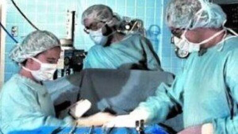
Innovative fluorescence-reading technology developed by researchers in the US will allow brain surgeons distinguish cancerous tissue from normal tissue.
The new tool, developed by the Norris Cotton Cancer Centre and Dartmouth College's Thayer School of Engineering, is already in use at the Cancer Centre and may become widely used around the world for surgeries treating a variety of cancers.
Removing a brain tumour is very intricate work for brain surgeons, and if any tumour tissue is left behind after the operation, it is likely that the tumour will regrow. Additionally, if they cut too much normal tissue the procedure can lead to permanent brain damage.
Keith Paulsen, PhD, a professor of biomedical engineering at Thayer School of Engineering and a member of the Cancer Imaging and Radiobiology Research Program at Norris Cotton Cancer Centre said: "Primary brain tumors look just like brain tissue.
"But if you look at them under a particular kind of light, they look much different."
The current method used is to give patients an oral dose of the chemical 5-aminolevulinic acid (ALA) which makes the tumour cells accumulate more PpIX- and therefore fluoresce, or "glow," when exposed to blue light. However, this method has been criticised for not being sensitive enough to highlight brain tumors that are less metabolically active, like low-grade gliomas.
In order to overcome this issue, MD-PhD student Pablo Valdes, PhD, and his research mentors-David Roberts, MD, the chief of neurosurgery at Dartmouth-Hitchcock Medical Center, and Dr. Paulsen used a probe that combines violet-blue and white light to simultaneously analyze both the concentration of PpIX and four other tumour biomarkers.
The probe can read how light travels when it hits the tissue, and sends this data to the computer which runs it through an algorithm and produces a straightforward answer as to whether the tissue is cancerous.
Dr. Paulsen said: "Our big discovery is that we can use the probe's reading of the fluorescing agent to measure the existence of a low-grade tumor in tissue.
"The probe is basically an enabling technology to show that information to the surgeon - a visual aid."
Posted by Fiona Griffiths