Columns (GC)
Published over 7 years ago. See the latest and most current information on Columns (GC).
Monoclonal antibodies (mAbs) have become increasingly important in the pharmaceutical industry with sales reaching $100 billion in 2017. This revenue is generated by blockbuster drugs such as Avastin, Herceptin or Remicade, targeting cancer or autoimmune diseases. In contrast to low-molecular weight drugs, the structural complexity of antibodies drives state-of-the-art chromatography and mass spectrometry (MS) to its limits. Either when seeking approval for new antibodies, or when developing biosimilars for products with an expiring patent, it is extremely important to be able to characterise these molecules in great detail.
Combining expertise from the analytical chromatography and semiconductor chip manufacturing industries, PharmaFluidics has created a new type of nano-LC column that is extremely well suited to handle these challenges. This new chromatographic column, called µPACTM, was devised based on in silico insights demonstrating the importance of order on the efficiency of chromatographic separations. Legacy LC columns contain randomly packed beads as their stationary phase. In contrast, PharmaFluidics uses a lithographic etching process to create a perfectly ordered separation bed on a silicon wafer.
Thanks to the perfectly ordered structure of the column, axial peak dispersion is virtually eliminated, resulting in ultra-high column plate numbers, narrower peaks and higher solute concentration in the peak maximum. The freestanding nature of the pillars also leads to much lower backpressure allowing the use of very long columns. These exceptional properties result in excellent chromatographic performance with high resolution and high sensitivity. This new approach significantly improves LC analysis of complex biological samples.
µPACTM’s micromachined separation bed is rigid and perfectly symmetrical. This allows it to be operated in both directions without risking stationary phase compression, leaching or column deterioration. The production process is highly reproducible, making each column completely identical. This makes µPACTM a robust high-resolution separation tool.
Discovered in the 1970’s, mAbs represented a major medical breakthrough. In the following decades, their therapeutic potential was unraveled, and biologics were created targeting a myriad of conditions. Currently, over 70 mAbs have received regulatory approval in the US and Europe. Over 20 of these have blockbuster status, generating billions of revenue every year. As many of these blockbuster drugs are close to coming out of patent protection, the creation of generics - called biosimilars - is on the rise. The first mAb biosimilars were approved in 2013 in Europe and 2016 in the US, and the number of applications is growing steadily since.
The specificity of mAbs was also leveraged to enable a more targeted application of cytotoxic drugs. This is achieved by linking such drugs to a mAb via a stable linker. The resulting compounds - called antibody-drug conjugates (ADC) - hold the promise of substantially lowering side effects of chemotherapy treatments. Currently, two ADC are already available to patients and over 30 are being evaluated for their safety and efficacy.
The ability to intensely characterise mAbs and ADC is essential, both for the production of new compounds and the approval of biosimilars. However, compared to small-molecule drugs, this is quite a challenging task. Monoclonal antibodies have a high molecular weight (ca.150 kDa) and are heterogenous in nature. Protein production in eukaryotic host cells leads to the introduction of many different post-translational modifications to the molecule. Furthermore, unwanted mutations in the host cell line can lead to an altered amino acid sequence and variations in higher order structures can also arise. In ADC, the structural variability resulting from the conjugation further increases the complexity. These properties greatly impact the safety and efficacy of the product and should thus be meticulously identified.
A powerful tool to obtain insight in the structure of mAbs and ADC is peptide mapping. Here, the large molecules are cleaved enzymatically into smaller elements, which can be separated by LC and identified by MS. The amount of detail that can be obtained from such an analysis is sufficient to determine the amino acid sequence and post-translational modifications. This enables its use in demonstrating similarity, for example between a biosimilar and its originator. Furthermore, drug conjugation sites can also be identified from the generated peptide map, extending its applicability to ADC.
Ever since its application in the characterisation of the first monoclonal antibodies in the late 1980’s, peptide mapping results have become more detailed thanks to the continuous technical improvement of the available equipment. A recent addition to the chromatographer’s toolbox is micro-pillar array columns. Their unique properties further improve resolution and sensitivity compared to the state-of-the-art conventional columns. This provides the performance needed to characterise mAbs in greater detail, as demonstrated in the following use cases.
The similarity of Herceptin (trastuzumab) and a candidate biosimilar was investigated by peptide mapping using µPACTM. Herceptin is a blockbuster mAb used in the treatment of HER2 positive breast cancer. As its patent protection is ending in 2019, many biosimilars are under development, hoping to claim a piece of the $6.6 billion market. Thus, powerful analytics are needed to evaluate their similarity is as much detail as possible.
Prior to the analysis, Herceptin was cleaved with trypsin. This enzyme’s specificity results in cleavage next to arginine and lysine residues. Based on the amino acid sequence of Herceptin, a total of 62 peptides would be expected. However, incomplete and non-specific cleavages drive up the number of peptides that is actually present. Furthermore, post-translational modifications on some of these peptides alter their physicochemical properties. Thus, over 100 peptides in greatly varying concentrations should be detectable from the sample to ensure a detailed characterisation. This drives chromatographic resolution to its limits.
Using µPACTM, 95% of the protein’s sequence could be mapped, including the identification of post-translational modifications. The remaining 5% of the sequence was represented in short fragments, which were not sufficiently retained on the column and eluted in the column flow-through. Nevertheless, the obtained coverage was exceptionally high and enables a detailed comparison between different production batches, or between originator and biosimilar.
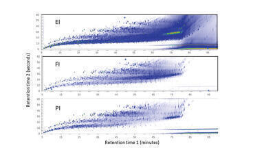
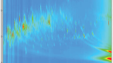
The results of such a comparison are shown in Figure 1. Highly comparable peptide maps were obtained for both Herceptin and the candidate biosimilar. However, the extracted ion chromatograms, shown in Figure 2, demonstrate that differences in post-translational modifications could be detected.
Another blockbuster mAb for which biosimilars have been created, and more are actively being pursued, is Remicade (infliximab). It is used in the treatment of autoimmune diseases and totalled $8.4 billion in global sales in 2015. Through peptide mapping with µPACTM, the similarity between Remicade and one of the biosimilars under development was evaluated.
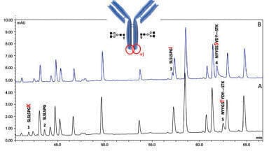
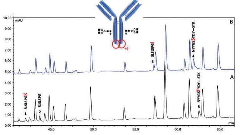
Following tryptic digest, the resulting peptides were separated on a µPACTM and detected via UV and MS (Figure 3). The chromatograms of both compounds are very similar, as expected, with the exception of a small number of peaks (1 - 5).
The origin of these discrepancies becomes clear from the MS data. Peaks 1 and 2 of the originator chromatogram are found to be absent for the biosimilar. They correspond to the peptides SLSLSPGK and SLSLSPG respectively, found in the C-terminal heavy chain of Herceptin. The variable presence of lysine at the end of the peptide is caused by an incomplete reaction of host cell carboxypeptidases on the antibody in the host cell during production. While these peaks are absent in the biosimilar chromatogram, an additional peak (3) appears, corresponding to the peptide SLSLSPGI. Thus, a point mutation in the biosimilar sequence is demonstrated. The two peaks in the Herceptin chromatogram are thus replaced by a single peak as there is no longer a lysine present at the C-terminal end that can be cleaved off by the host-cell carboxypeptidase.
Similarly, an additional point mutation was discovered as seen in the different retention time of peaks 4 and 5. From the MS/MS data shown in Figure 4, it becomes apparent this corresponds to a threonine to serine amino acid substitution.
The discovery of these differences in primary sequence is of great significance, as this by definition rules the candidate biosimilar out from further development in both the US and Europe.
Created by conjugating Herceptin with the cytotoxic drug DM1, Kadcyla (ado-trastuzumab emtansine) is also applied in the treatment of HER2 positive breast cancer. Both molecules are chemically linked via a non-reducible thioether linker on the antibody’s lysine residues. With many lysine residues present throughout the Herceptin structure, a drug distribution of 0 - 8, and a drug-to-antibody ratio of 3.5, thousands of species can be generated during the conjugation process. Through peptide mapping with µPACTM, the drug conjugation sites could be determined.
Since the mAb part of Kadcyla corresponds exactly to Herceptin, a highly similar chromatogram is expected. The only differences would then originate from the conjugation with DM1. This is indeed what is seen from their LC-UV and MS total compound chromatograms presented in Figures 5 and 6. The new peaks present in the Kadcyla chromatogram elute later, which corresponds to the increased hydrophobicity introduced by the presence of DM1.
The conjugation of DM1 on these peptides is confirmed by their collision-induced dissociation (CID), where specific fragments from the cytotoxic drug arise. For example, the ion at m/z 547.2206 can be used to identify conjugated peptides in the LC-MS chromatogram. As shown in Figure 7, this small number of peaks in this chromatogram illustrates the selectivity of the all-ions MS/MS functionality.
Finally, Figure 8 shows the extracted ion chromatograms of a selection of identified conjugated peptides. These chromatograms surprisingly show two peaks per conjugated peptide, corresponding to two distinct stereochemical configurations of the linkage. This means that peptide mapping with µPAC™ not only reveals the conjugation sites themselves, but can also achieve the separation of diastereomers, providing additional information.
Micro pillar array columns are an important new addition to the chromatographer’s toolbox in the characterisation of monoclonal antibodies and antibody-drug conjugates. An excellent sequence coverage is obtained in peptide mapping experiments. Essential insights in the antibody’s sequence and structure are revealed, allowing a detailed comparison between different production batches or between originators and candidate biosimilars. The use cases described in this article demonstrated that µPACTM was capable of uncovering differences in post-translational modifications and revealing point mutations. Furthermore, in addition to the conjugation sites of ADC, the stereochemical configurations of these conjugations could be defined. This shows that µPAC™ is well equipped to handle the analytical challenges that are associated with these complex molecules.
We would like to thank Dr Koen Sandra, Scientific Director of the RIC (Research Institute of Chromatography), Belgium, for his input and contribution.
[1] Sandra et al. (2018). LCGC Europe, 31(3), 155-166