Bioanalytical
Published over 4 years ago. See the latest and most current information on Bioanalytical.
Plasma is a common matrix used in bioanalysis. However, the phospholipids present in plasma can cause a range of issues with analysis such as higher backpressures, an increased need for instrument maintenance and matrix effects in the form of ion suppression when using ESI-MS detection. Ion suppression leads to other issues during chromatographic runs such as irreproducible results, loss of signal intensity and poor quantitation. This application note explores the use of the Microlute® PLR 96 well plate to show it is possible to achieve highly reproducible recoveries alongside removal of phospholipids to eliminate the problems they cause with analysis.
Blood plasma is an important matrix in drug metabolism and pharmacokinetic (DMPK) studies where it is used in large quantities for bioanalysis work [1]. For drug development in DMPK, different compounds are screened and properties assessed, this is usually by Liquid Chromatography - Mass Spectrometry (LC-MS) [2]. In order for LC-MS to be used for drug development analysis, it requires sample clean-up to remove interferences from plasma before injection onto the system to obtain reliable and consistent results.
Clean-up of plasma can be performed before analysis in several different ways. The simplest preparation method is to use protein precipitation. This process involves adding an organic solvent, ‘salting out’ or by adjusting the pH of the plasma solution which then causes the proteins to crash out of solution. It is a quick and easy method to prepare the solution and prevent the proteins from affecting downstream applications when injected onto the chromatography instrument. However, this preparation technique does not remove phospholipids from the sample which can cause a number of complications and hinder results.
Phospholipids are a class of lipids which contains both a phosphate head and up to two fatty acid derived tails. This results in them being amphipathic - the polar phosphate head giving the hydrophilic property with the tails responsible for hydrophobic properties. Phospholipids are a major component of cell membranes are thus ubiquitously found throughout the human body. The most common classes of phospholipids are the glycerophospholipids (which are composed primarily of glycerophosphocholines), lysophospholipids and sphingolipids (Figure 1) which make up 70% (glycerophospholipids), 10% (lysophospholipids) and between 10 - 20% (sphingolipids) of total phospholipids [3,4].
Of these phospholipids, there are several sub-classes of common phospholipids - phosphatidic acid (PA), phosphatidylcholine (PC), phosphatidylethanolamine (PE) and phosphatidylserine (PS) and sphingomyelin (Figure 2).
It is known that reversed phase columns, commonly used in LC-MS methods (e.g. C8 and C18), suffer from reduced column lifetime from phospholipids in the sample that are injected onto the column [5]. This occurs when the hydrophobic functional groups on the column interact with the hydrophobic tails of the phospholipids. Typically, some phospholipids could become bound onto the column’s stationary phase resulting in reduced column capacity for retention, higher system back pressures and slowly bleeding off the column at random intervals. It is possible to include in a high organic flush to prevent this, but this can add on time to already long analysis method times [6].
Phospholipids are also known to cause ion suppression. This occurs in the mass spectrometer source when an interfering compound co-elutes from the HPLC column at the same time as an analyte which is being analysed. A co-eluted molecule, such as a phospholipid, can compete for the available charge in the source with the analyte of interest causing less of the analyte to be ionised. This reduces the intensity of the analyte and hence signal suppression occurs. Phospholipids can also influence evaporation of the solvent droplets entering the source. Reducing the volatility of the droplet may increase the radius and reduce surface area and charge thereby, reducing the overall intensity of the signal for the analyte [3, 7].
Solid phase extraction (SPE) is a popular method for removing phospholipids when preparing plasma samples. Wash steps remove interfering compounds from samples and allow elution of cleaner solutions ready to be injected straight onto the system of choice or be dried down and reconstituted into a suitable solvent mix. Although SPE has its advantages, it can be time-consuming to develop a protocol which works effectively and reproducibly to remove both proteins and phospholipids.
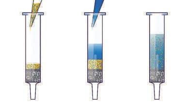
The Microlute® PLR product is specifically designed to remove protein and phospholipids from plasma prior to LC-MS analysis. It provides a quicker and easier method to efficiently remove proteins and phospholipids from plasma and prepare samples for injection (Figure 3).
The advantage of this method over other sample preparation techniques is the speed and ease of developing a method to run plasma samples. Method development is as simple as either diluting viscous samples with water or adjusting the volume of organic solvent at the protein crash step. Compared to SPE, phospholipid removal by Microlute® PLR offers a faster and simpler process to develop methods allowing higher throughput with less hands-on time.
This application explores the differences of phospholipids present in human, bovine, pig and rat plasma sources, specifically the composition of phospholipids present within each type. Along with analysis of analyte recovery and removal of each of the different phospholipids using the Microlute® PLR 96 well plate (PPLR025P-001). These sources are from the three classes commonly used in DMPK studies - human, rodent (rat) and non-rodent (bovine and pig). Here we demonstrate that despite different sources of plasma, reproducible recovery and phospholipid removal can be achieved.
Experimental
The compounds tested in this application note consisted of two acidic, basic and neutral analytes. This tested the flexibility of the product to give good recovery results independent of the compound classification. There was a range of hydrophobicities between the compounds which spanned from 1.61 for hydrocortisone up to 4.92 for amitriptyline to test the impact of hydrophobicity of the analytes on recovery.
Sample Preparation Method
Human, pig, bovine and rat plasma were chosen for this application. All plasma was obtained from Sigma-Aldrich®.
For each of the different plasma types, a volume of plasma was spiked to a concentration of 1.5 µg/mL and allowed to equilibrate for 30 minutes. 100 µL of each of the spiked plasma samples was added to six wells of the plate and to just a single well, 100 µL of each type of blank plasma was added for matrix match standards. All plasma samples were crashed with 300 µL of an acetonitrile solution containing 1% formic acid and aspirated four times with a pipette to ensure that the two solutions were completely mixed. The crashed plasma solution was eluted into a 1 mL collection plate (Cat no. 219250) using 5 PSI of positive pressure until the wells were completely empty. The solutions prepared in the collection plate were injected directly onto the LC-MS system and analysed using the conditions set out in Tables 2 and 3. Each of the matrix match standards were spiked to a concentration of 0.375 µg/mL.
Results and Discussion
Chromatography
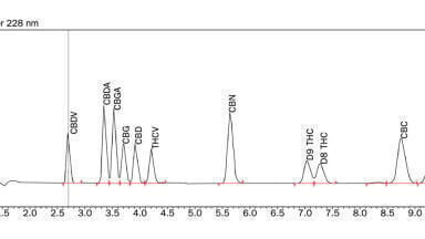
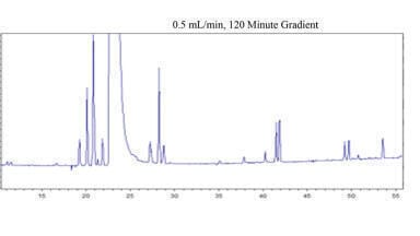
Figure 4 shows the chromatography for the method showing only the analyte peaks. There are no major interfering peaks present throughout. The early eluting peaks do tail slightly which is due to the 75% acetonitrile solution being injected from the collection plate. This could be improved by diluting with water or drying down and reconstituting in a lower elution strength solution. For the purpose of this application, this was not performed as the LC method showed good reproducibility injection-to-injection and demonstrated the ability to quickly crash, elute and inject onto a system and obtain high and reproducible recovery.
Figure 5 shows the phospholipid peaks from the method using the protein precipitated samples. The peaks separated out well, with the majority having no interferences. The pig plasma has a shoulder peak on the 34:1 SM peak that is likely due to the presence of an isomer. Due to the single quadrupole instrument used, it is not possible to separate out these isomers.
Plasma Comparison
This application note focused on six different phospholipids which are common in plasma - 16:0 LPC, 18:0 LPC, 34:1 SM, 34:2 PC, 36:2 PC and 38:4 PC. These range from a LogP of 5.6 for 16:0 LPC to 14.1 for 38:4 PC [9]. The shorter chained phospholipids (16:0 and 18:0 LPC) are most likely to co-elute with analytes due to their lower hydrophobicity. The longer chained phospholipids are more likely to stick to the HPLC columns and/or elute at irregular intervals and cause unexpected peaks and matrix effects.
For this application it was useful to show the relative difference of the amount of each phospholipid present in each sample. Table 4 shows the different relative compositions of the phospholipids in each protein precipitate plasma sample with the values plotted for comparison in Figure 6. From these values, it is possible to compare the compositions of each of the different plasma samples to each other.
This is a useful indication of how easy or difficult it would be to remove the different phospholipids, their likelihood in interfering with ionisation or whether they are more likely to cause issues with build-up on LC columns. For example, a higher amount of the less hydrophobic phospholipids (16:0 or 18:0 LPC) would indicate it is less likely to cause LC column build-up but more likely to end up eluting with analytes. While the inverse case of higher amounts of hydrophobic phospholipids would potentially end up with them being bound to the LC column.
Phospholipid Removal
Phospholipid removal is an important consideration with bioanalysis due to the advantages that could be gained [12]:
• It leads to more reproducible methods due to matrix effects being reduced. Reproducible methods mean less samples need to be tested to be confident in results analysed.
• More sensitive methods due to reduced ion suppression. This allows for less sample to be used during analysis.
• Increased column lifetime by reducing phospholipids being adsorbed onto the stationary phase.
• Reducing contaminating matrix components entering the source to minimise maintenance downtime.
Table 5 shows the phospholipid removal for each of the plasma samples tested. All highly hydrophobic phospholipids were removed. For the two lysophospholipids, only a trace amount were left with the lowest removal of 99.4% for both the pig and human plasma. The total phospholipid removal was calculated by weighting each removal by the composition of the phospholipids analysed in Table 4. This data showed that phospholipid removal was achieved for each of the four samples.
Recovery
Recovery of analytes from each plasma processed with the Microlute® PLR 96 well plate is shown in Figure 7. Each value plotted is the mean recovery of six replicates injected onto the LC-MS system. The analytes were a mixture of acidic, basic and neutral compounds over a range of hydrophobicities - see Table 1 for details on the compounds. All recoveries were greater than 90% showing the Microlute® PLR is highly effective and reproducible for obtaining high recoveries without compromising the removal of phospholipids.
Reproducibility
Well-to-well reproducibility is an important metric to provide confidence in data. The lower the %RSD value, the more reproducible the results. Chromatographic bioanalytical methods typically have a guideline of 10-15 % RSD limit set when the method is validated [13, 14].
Figure 8 shows the data for the reproducibility. The data ranged from 0.88 %RSD for amitriptyline in bovine plasma to 5.74% RSD for ketoprofen in pig plasma. The data show that the reproducibility for every analyte in the four plasmas were well in specification to meet the typical %RSD guidelines chosen during bioanalytical method validation for drug development.
Summary
Microlute® PLR 96 well plate is an easy-to-use product with a quick protocol. It offers complete phospholipid removal for a range of plasma samples with differing phospholipid compositions and properties (viscosity and protein amount) with no compromise on recovery or reproducibility of analytes.
References:
1. W. Wang, J. Liu, Y. Han, W. Huang and Q. Wang, “The most convenient and general approach for plasma sample clean-up: multifunction adsorption and supported liquid extraction,” Bioanalysis, vol. 4, no. 3, pp. 223-225, 2012.
2. Korfmacher, “Mass Spectrometry: The Premier Analytical Tool for DMPK Scientists in a Drug Discovery Environment,” LCGC North America, pp. 640-647, 1 August 2012.
3. E. Chambers, D. M. Wagrowski-Diehl and J. R. Mazzeo, “Systematic and comprehensive strategy for reducing matrix effects in LC/MS/MS analyses,” Journal of Chromatography B, vol. 852, no. 1-2, pp. 22-34, 2007.
4. M. Koval and R. E. Pagano, “Intracellular transport and metabolism of sphingomyelin,” Biochimica et Biophysica Acta, vol. 1082, no. 2, pp. 113-125, 1991.
5. J. Carmical and S. Brown, “The impact of phospholipids and phospholipid removal on bioanalytical method performance,” Biomedical Chromatography, vol. 30, no. 5, pp. 710-720, 2015.
6. D. Neville, R. Houghton and S. Garrett, “Efficacy of plasma phospholipid removal during sample preparation and subsequent retention under typical UHPLC conditions,” Bioanalysis, vol. 4, no. 7, pp. 795-807, 2021.
7. O. A. Ismaiel, M. S. Halquist, M. Y. Elmamly, A. Shalaby and H. T. Karnes, “Monitoring phospholipids for assessment of ion enhancement and ion suppression in ESI and APCI LC/MS/MS for chlorpheniramine in human plasma and the importance of multiple source matrix effect evaluations,” Journal of Chromatography B, vol. 875, no. 2, pp. 333-343, 2008.
8. “Drugbank,” Drugbank, [Online]. Available: https://go.drugbank.com/. [Accessed 2021].
9. “PubChem,” National Library of Medicine, [Online]. Available: https://pubchem.ncbi.nlm.nih.gov/. [Accessed 2021].
10. U. Windberger, A. Bartholovitsch, R. Plasenzotti, J. Korak and G. Heinze, “Whole blood viscosity, plasma viscosity and erythrocyte aggregation in nine mammalian species: reference values and comparison on data,” Experimental Physiology, vol. 88, no. 3, pp. 431-440, 2004.
11. A. Swan, G. T. Allen and N. C. Tanner, “The blood volume and plasma protein levels,” Gut, vol. 3, p. 149, 1962.
12. J. Danaceau, H. Yu, E. Chambers and K. J. Fountain, “Matrix effects in metabolite quantification for MIST assessment: the impact of phospholipid removal and HPLC column particle size,” Bioanalysis, vol. 6, no. 6, pp. 761-771, 2014.
13. Food and Drug Administration (FDA), 2018. [Online]. Available: https://www.fda.gov/files/drugs/published/Bioanalytical-Method-Validation-Guidance-for-Industry.pdf.
14. European Medicines Agency (EMA), 2019. [Online]. Available: https://www.ema.europa.eu/en/documents/scientific-guideline/draft-ich-guideline-m10-bioanalytical-method-validation-step-2b_en.pdf.