Electrophoretic separations
Published over 14 years ago. See the latest and most current information on Electrophoretic separations.
Researchers have attempted to fill in gaps in the adult development during the non-feeding stage by analysing detailed protein structural information for individual PPFB and PVFB tissues during larval and pupal developmental stages.
Previous research has established that peripheral fat body (PPFB) tissues and pupal perivisceral fat body (PVFB) tissues act anti-apoptotic besides being assigned roles in embryogenesis and defense. However, detailed protein structural information for individual PPFB and PVFB tissues during larval and pupal developmental stages is still missing.
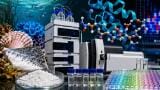
Using gel electrophoresis and chromatography to separate the 30 kDa proteins from both PPFB and PVFB as well as hemolymph total proteomes, as well as Mass spectrometry (MS) to elucidate individual protein sequences, the researchers found that 30 kDa lipoproteins LP1 to LP5 and L301/302 were detected in PPFB and PVFB tissue as well as in hemolymph in B. mori larvae and pupae.
They also observed that the concentration of the proteins changed progressively during development from their synthesis in PPFB, transport in hemolymph to storage in PVFB. The amino acid sequences of all known 30 kDa proteins showed very high homology.
Posted by Neil Clark