Electrophoretic separations
Published over 14 years ago. See the latest and most current information on Electrophoretic separations.
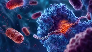
There are several ways in which researchers can explore the molecular imaging of tissue sections by mass spectrometry (MS), opening exciting avenues for exploration by producing molecular images directly from sections with high mass accuracy.
Time-of-flight secondary ion mass spectrometry (TOF-SIMS) is one such technique, and it has proved to be an accurate method for generating molecular images and depth profiles of samples, including tissue sections.
SIMS use high energy, high spatial resolution ion beam to bombard the sample surface resulting in the ejection of secondary ions originating from the molecules on the surface that can be measured by mass spectrometry. These can be either liquid metal ion guns (LMIG) or cluster guns, which both have their merits and drawbacks.
There are also two approaches to three dimensional SIMS imaging. One method is collecting serial sections of a tissue and individual 2D images which are later reconstructed into a 3D image. The second method is a depth profiling approach in which a single biological sample is eroded and imaged generating several layers of mass spectral data throughout a z-stack of the sample.
The technology has the ability to generate chemical images of surfaces with high sensitivities and minimal chemical damage, and new technologies are consistently evolving allowing for greater accuracy and reliability.
Posted by Fiona Griffiths