Preparative
Published over 9 years ago. See the latest and most current information on Preparative.
Antibody drug conjugates (ADCs) represent an exciting and rapidly growing class of biotherapeutics. Because of their unique molecular structure, pharmacokinetic studies of ADCs can be particularly challenging. Ligand binding assays (LBA) have typically been used but can suffer from poor reproducibility, limited dynamic range, and cross reactivity. Liquid chromatography tandem mass spectrometry (LC-MS/MS) assays generally have high selectivity, dynamic range, and reproducibility but can lack in sensitivity when applied to protein therapeutics. In this article, we describe a hybrid LBA/LC-MS/MS technique that uses a universal immunoenrichment strategy and microflow LC to create a novel quantitative analysis solution that accelerates method development and improves performance of ADC pharmacokinetic assays.
Within the last several decades, monoclonal antibody (mAb) based drugs have become an established class of biotherapeutics and represent the fastest growing segment of the global biopharmaceutical market [1-3]. Because of their inherent advantages in specificity, efficacy, and safety, pharmaceutical companies have dedicated large portions of their research and development efforts to mAb-based therapies with the list of approved and investigational mAb based drugs now reaching into the hundreds [4].
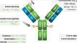
MAb based therapies represent a way to specifically target a disease by using an immune molecule which, by design only targets the diseased cells of interest. Antibody drug conjugates (ADCs) are a class of biotherapeutics that combine a mAb with a cytotoxic small molecule drug attached to the mAb through a chemical linker group. The idea behind an ADC is that the antibody provides the specificity while the small molecule drug provides the cytotoxic payload to treat the disease. This targeted delivery of the cytotoxic drug improves the drug’s overall efficacy and minimises systemic toxicity.
The unique chemical structure of ADCs, as both an antibody and a small molecule drug, can present challenges for scientists tasked with their bioanalysis. Most ADCs are heterogeneous molecules. One or more drugs may attach to the antibody at various sites resulting in varying drug to antibody ratios (DAR). To add to the complexity, the chemical lability of the linker group as well as the chemistry used for conjugation can affect the in vivo stability and heterogeneity of the ADC. Thus, it can be a challenge to determine which form or forms of the ADC molecule should be monitored.
Traditionally, ligand binding assays (LBA) such as enzyme linked immunosorbent assays (ELISA) have been used for the bioanalysis of mAbs and mAb based biotherapeutics. LBAs typically have high sensitivity and high throughput, are relatively inexpensive, and require limited sample preparation. However, LBAs can suffer from high variability, narrow dynamic range, and problems with selectivity. Additionally, internal standards cannot be added which help to correct for anomalies and ensure accurate quantification.
Mass spectrometry, and in particular liquid chromatography tandem mass spectrometry (LC-MS/MS) has found widespread use for the quantitative analysis of small molecule drugs. Assays based on LC-MS/MS are exceedingly sensitive and selective with excellent dynamic range. Additionally, assays can be multiplexed and internal standards are easily added to any experiment. In more recent years LC-MS/MS has been applied to the bioanalysis of mAbs and other proteins in biological fluids. Because of their large size, proteins are typically not quantified intact but instead are digested into peptides, with one or more peptides chosen as a proxy for quantification of the protein. However, quantification of mAbs and other proteins from plasma often requires greater sensitivity than can be achieved with LC-MS/MS alone using traditional LC flow rates. Additionally, as potency increases, dosage is decreased, thereby resulting in less biotherapeutic in circulation to measure.
Here we describe a universal immunocapture enrichment strategy coupled with sample preparation and microflow LC-MS/MS analysis and apply it to the total antibody analysis of the ADC ado-trastuzumab emtansine in plasma. Streptavidin coated magnetic beads bound with a universal capture reagent accelerate method development for total antibody enrichment. The use of microflow LC provides greater sensitivity than traditional flow LC. The hybrid LBA microflow LC-MS/MS workflow provides a customisable immunocapture strategy that enables the rapid development of high sensitivity pharmacokinetic assays of biotherapeutics during pre-clinical or phase I-IV studies.
Magnetic Bead Preparation: Magnetic immunocapture beads from the BioBA sample prep kit (Sciex) were prepared with biotinylated goat anti-human IgG antibody (included in the kit) according to the kit protocol.
Sample Preparation: 10x spiking solutions of ado-trastuzumab emtansine were first prepared in 1X BioBA bind/wash buffer containing 0.01% BSA (bovine serum albumin), then spiked into Sprague-Dawley rat plasma, K2EDTA (BioreclamationIVT) at the final concentrations of 0.5-100,000 ng/ml. SILuMab (Sigma Aldrich), was used as internal standard (IS) and was added to the plasma samples prior to BioBA immunocapture processing. A blank and double blank sample were also prepared. The double blank only had 1X BioBA bind/wash buffer containing 0.01% BSA and the blank sample had rat plasma with additional internal standard. Spiked plasma samples (50 µL) were mixed with internal standard and processed based on the BioBA generic method with some modified steps to reduce background signal and improve signal to noise (S/N) ratio in order to utilise the advantage of higher sensitivity provided by microflow LC. The modified steps include: an extra hour of incubation of conjugated beads with sample followed with two wash steps with 500 µL of BioBA bind/wash buffer and 500 µL of 50 mM ammonium bicarbonate. Each immunopurified sample was digested with 1 µg total Trypsin/Lys-C.
Traditional Flow Liquid Chromatography: A Shimadzu Prominence HPLC system with two LC-20AD pumps, CTO-20A column oven, and a SIL-20AC autosampler was used. The column was a 100 x 2.1 mm Kinetex C18 2.6 µm 100 Å column (Phenomenex). Mobile phase A, water with 0.1% formic acid, and mobile phase B, acetonitrile with 0.1% formic acid, was used at a flow rate of 0.5 ml/min. Wash solvent for the autosampler was 20/20/60 methanol/acetonitrile/IPA. Injection volume was 25 µL, and the column was kept at 40°C. The gradient method was as follows: 0 min, 5% B; 0.7 min, 5% B; 0.8 min, 10% B; 3.5 min, 25% B; 5.0 min, 40% B; 5.1 min, 95% B; 5.9 min, 95% B; 6.0 min, 3% B; 7.0 min, 3% B.
Microflow Liquid Chromatography: A Sciex M3 MicroLC-TE system, with two microLC gradient pumps and an integrated autosampler was used in combination with a source mounted column oven (Sciex). A 10 x 0.3 mm trap column packed with 5 µm 120 Å ChromXP C18 CL and an analytical column 50 x 0.3 mm HALO Peptide ES-C18 2.7 µm 160 Å column was used (Sciex). Mobile phase A in the analytical gradient was water with 0.1% formic acid, mobile phase B was acetonitrile with 0.1% formic acid with flowrate of 10 µL/min. The column temperature was set to 40°C. Injection volume was 25 µL, and the autosampler needle and valve wash consisted of two cycles using mobile phase B, followed by one cycle using mobile phase A. The gradient method was as follows: 0 min, 3% B; 0.7 min, 5% B; 0.8 min, 10% B; 3.5 min, 25% B; 5.0 min, 40% B; 5.1 min, 95% B; 10.0 min, 95% B; 10.1 min, 3% B; 15.0 min, 3% B. For trapping conditions, mobile phase A in the loading gradient was water with 0.1% formic acid, mobile phase B was acetonitrile with 0.1% formic acid. Sample was loaded from the injection loop onto the trap column using 100% A for 2.5 min at 50 µL/min flow rate. The trap was then washed with 95% B followed by 100% A each at 50 µL/min for 5 min after every injection.
Mass Spectrometry and Data Processing: A Sciex QTRAP® 6500+ with IonDrive™ Turbo V source was used. For the microflow LC experiments, the standard electrode was replaced with a 25 µm ID electrode (Sciex). The transitions and MS parameters were optimised using DiscoveryQuant™ software (Sciex) and kept constant for both the traditional flow and microflow LC experiments. MultiQuant™ 3.0.2 software (Sciex) was used for data analysis. Sample for both microflow and traditional flow LC-MS/MS analysis was prepared on the same day to exclude variations in response due to sample preparation. Three replicate LC-MS/MS injections were acquired for both the traditional flow and trap-and elute microflow LC analysis.
Ado-trastuzumab emtansine is the first HER2-targeted treatment for metastatic breast cancer. It combines the mAb trastuzumab with the cytotoxic agent emtansine bound through a non-cleavable chemical linker to lysine groups on the mAb. Due to the availability of multiple lysine sites for emtansine conjugation, the final drug product is a heterogeneous mixture containing various numbers of emtansine molecules. This results in various drug to antibody ratios (DAR) from 1 to 8 in the final product. In order to accurately quantify all species of the drug product, several bioanalytical assays are required.
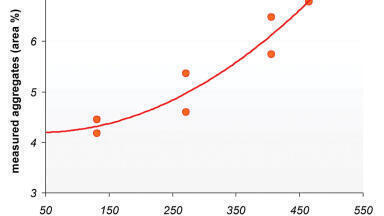
The high capacity BioBA sample enrichment kit used in this study utilises magnetic beads to facilitate capture and recovery of samples from complex matrices such as plasma. The BioBA magnetic beads are cellulose based particles with a macroporous structure and are on average 30-50 µm in size. The hydrophilic cellulose surface and macroporous structure yield a particle with low nonspecific binding and higher surface area than polymeric particles. Magnetic beads offer several advantages including: ease of handling, scalability, improved sample recovery, parallel processing of samples using a variety of magnetic stands, and use in high-throughput formats with robotics. A study on the binding capacity of the BioBA beads using biotinylated anti-human IgG and monitoring its disappearance from the supernatant over time using a bicinchoninic assay showed that binding was complete (96%) after 60 minutes (Figure 1).
The beads have high binding capacity with 1 mg of beads capable of binding 77 μg of biotinylated anti-human IgG, allowing for capture of more target analyte proteins than other polymer based commercially available magnetic beads (Figure 2).
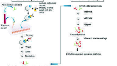
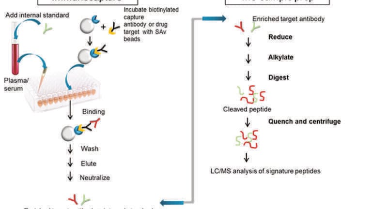
The magnetic beads in the BioBA kit are coated with streptavidin which has an extremely high affinity for biotin. This enables an easily customisable immunocapture strategy. Various LBA LC-MS/MS assays can be developed by first binding the appropriate biotin conjugated immunocapture reagent to the streptavidin coated beads. For example, for heterogeneous ADC drug products such as ado-trastuzumab emtansine, the amount of conjugated ADC species can be determined using an anti-payload antibody for immunocapture. To assay the total antibody (free + conjugated), a generic anti-human Fc antibody can be employed for immunocapture, or a target specific immunocapture strategy can be employed with recombinant target protein or an anti-idiotype antibody. For the current study, an assay was created to determine the total antibody (DAR 0 to 8) in the drug product by binding biotinylated goat anti-human IgG antibody to the beads as the immunocapture reagent. This antibody is included in the kit and provides a universal means to capture human mAbs. It will capture all ADC species including those without payload attached and provides a total antibody measurement. After capture and elution from the beads, samples are reduced, alkylated, and digested with a Trypsin/Lys C enzyme combination using reagents and buffers included in the BioBA kit which includes a mass spec compatible anionic surfactant (Figure 3). Because the samples also contain the SILuMab internal standard (IS), this protein is also digested. The digestion efficiency of the IS does not need to be equivalent to the ADC as long as it is reproducible.
Once the protein has been digested, one or more peptides are used as surrogates for protein quantification. The ado-trastuzumab emtansine conjugation is through lysine groups but in general, lysine containing peptides should be avoided as their cleavage may not be reproducible. The signature peptides IYPTNGYTR and FTISADTSK from the CDR region and the conserved Fc peptide DTLMISR are typically chosen for quantification of trastuzumab. However, since the peptide FTISADTSK contains a lysine, the two signature peptides IYPTNGYTR and DTLMISR are preferred for ado-trastuzumab emtansine. Peptide IYPTNGYTR is a unique peptide to trastuzumab from the CDR region, peptide DTLMISR is a universally conserved human IgG peptide. The SILuMab internal standard (IS) yields a heavy labelled peptide for DTLMISR. This heavy labelled peptide was used as the IS for all signature peptides used for quantification as it is very close in retention time to the analyte peptides.
The most effective LC-MS/MS assays for quantification use a technique called multiple reaction monitoring, or MRM. In MRM, the precursor ion of a specific analyte is selected in the first mass analyser, fragmented in a collision cell, and then one or more of the resulting product ions are monitored in the second mass analyser. This provides a uniquely selective and sensitive method for quantification. For the signature peptides chosen here, DiscoveryQuant™ software was used to aid in the selection and optimisation of the best product ions for each peptide precursor. The software will automatically ramp MS parameters in a series of experiments to find the best MS conditions and fragmentation energies for each MRM transition, greatly expediting the development of the final assay.
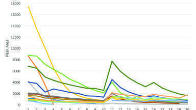
Figure 4 shows the extracted ion chromatograms (XICs) of the signature peptide FTISADTSK at the 5 ng/ml and 10 ug/ml level for both traditional flow and microflow LC. The S/N ratio was improved by 4 fold for this signature peptide as well as IYPTNGYTR using microflow LC.
Table 1 shows the comparison of the quantification statistics generated using MultiQuant Software for data acquired by traditional and microflow LC for the signature peptide IYPTNGYTR. Figure 5 shows the calibration curve for this same peptide using Microflow LC. The LLOQs for both methods were determined using the requirements of precision < 20% and accuracy between 80 and 120% at LLOQ, and at any higher concentration a precision < 15% and accuracy between 85% and 115%. LLOQ improved by a factor of 5 using the microflow LC trap-and-elute method using both signature peptides and the conserved Fc peptide DTLMISR. For the traditional LC method the limit of quantification LOQ was 5 ng/ml whereas an LOQ of 1 ng/ml was achieved by microflow LC. Both traditional flow and microflow LC methods showed good linearity with r > 0.99.
A universal LBA/LC-MS/MS workflow using high capacity streptavidin coated magnetic beads and reagents supplied with the BioBA sample prep kit enable the rapid development of pharmacokinetic assays of antibody based therapeutics. The versatility of the immunoenrichment strategy enables customisation by binding the appropriate biotin conjugated immunocapture reagent to the streptavidin coated beads. As shown here, the binding of the magnetic beads with a universal capture reagent followed by LC-MS/MS analysis enabled the accelerated development of the total antibody assay for ado-trastuzumab emtansine with wide dynamic range, high selectivity, and high sensitivity, with an LLOQ at low nanogram levels. Microflow LC enabled up to 5x lower LLOQ vs. traditional flow LC providing a solution for applications where quantification is needed at low concentrations and/or when sample volumes are limited. The workflow is generally applicable to mAb based therapeutics and results in faster assay development with more selectivity and lower LLOQs than typically achieved with LC-MS/MS using a direct plasma or pellet digest.
[1] X. Geng, X. Kong, H. Hu, J. Chen, F. Yang, H. Liang, X. Chen, and Y. Hu, Hum Vaccin Immunother. 11(12) (2015) 2769–2776.
[2] D. Kuystermans and M. Al-Rubeai in M. Al-Rubeai (Editor), Animal Cell Culture, Springer, Dordrecht, The Netherlands, 2015, 717-57.
[3] M. Stern, R. Herrmann, Crit Rev Oncol Hematol. 54 (2005) 11-29.
[4] https://en.wikipedia.org/wiki/List_of_therapeutic_monoclonal_antibodies