Preparative
Published over 5 years ago. See the latest and most current information on Preparative.
Imaged capillary isoelectric focusing (iCIEF) is a high throughput, highly efficient separation technique in routine use by many biopharmaceutical companies for the determination of the charge heterogeneity of proteins. Various charge variants detected in the sample limit the application of iCIEF as a powerful tool in multi-attribute methods. Characterisation of these variants is required
In this report, high-efficiency protein charge variant fractionation characterisation with a newly developed preparative iCIEF will be illustrated. High-efficiency iCIEF charge variant peak separation and collection, followed by mass spectrometry (MS) characterisation or online iCIEF-MS with the National Institute of standards and Technology Humanized IgG1κ Monoclonal Antibody (NISTmAb) will be demonstrated.
capillary isoelectric focusing, CIEF; critical quality attribute, CQA; imaged capillary isoelectric focusing, iCIEF; post-translational modification, PTM; multi attribute method, MAM; ion exchange chromatography, IEC; whole column image detection, WCID; electro-osmotic flow, EOF.
Analysing charge variants of therapeutic proteins is essential for the characterising and monitoring of critical quality attributes such as physicochemical and immunochemical properties, biological activity, and quantity during development and manufacturing. Protein charge variants arise by deamidation, formation of N-terminal pyroglutamate, aggregation, isomerisation, sialylation, antibody fragmentation, and glycation of lysine residues. These changes affect target binding, biological activity, patient safety, and shelf life [1-3].
Imaged capillary isoelectric focusing (iCIEF) has become an indispensable tool in therapeutic protein development and manufacturing because of its high analytical throughput, ease of use, fast method development, and excellent reproducibility. Compared to other charge-based separation techniques, iCIEF can usually separate protein charge variants in a fraction of the time required in contrast to a CIEF method on a conventional capillary electrophoresis instrument.
There is a tremendous and urgent need for the identification of these protein charge variants. Collection and subsequent characterisation of individual variants in a protein sample are critical for understanding the charge distribution’s nature on a molecular level [3,4]. The origin, stability and biological activity of charge variant is crucial during protein therapeutic discovery, formulation, stability study, regulatory agency approval, production, and quality control. This has been a challenge until the introduction of preparative iCIEF.
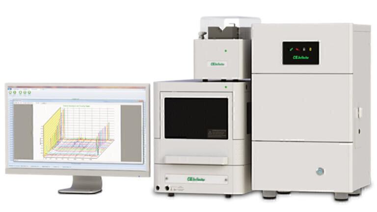
A generic iCIEF platform has been developed, CEInfinite (AES Ltd, Canada), on which methods for different protein samples are readily developed [2,3].
The amount and purity of charge variant isomers collected by the traditional slab gel IEF method is usually insufficient for further characterisation [3]. Fractionation and collection of sample zones in CIEF is unpractical on conventional CE instruments and commercially not available.
Nowadays, IEC is exploited for protein charge variant fractionation. Although both IEC and IEF are charge-based protein separation methods, different mechanisms are involved. As a result, protein charge variants may not correlate well between IEC to the IEF. Since IEC is a low-resolution separation, unsatisfactory purity and high salt content are usually expected of the IEC’s fractions. [3].
With the introduction and commercialisation of a preparative system iCIEF system, CEInfinite, shown in Figure 1, highly efficient fraction collection of protein charge variant according to protein iCIEF profile in µg to low mg amounts has become possible. Existing iCIEF methods can be easily transferred to prep iCIEF with minimal method development. With a patented 320 µm prep iCIEF cartridge [4], charge variants can be collected and can be used for intact MS analysis and peptide mapping characterisation in one day. The highly efficient prep iCIEF has been applied in therapeutic protein discovery, formulation study, new drug application, and routine MAM protein characterisation. The outstanding advantages of iCIEF in terms of simple method development, high-resolution separation, and robustness are powered by the capability of straightforward and high-efficiency capillary-based prep iCIEF.
MAM is a highly sought-after tool in biopharma for some time [5]. As a high-resolution charge-based separation and sample concentrating technique iCIEF is the most powerful unequivocal molecular weight structure characterisation method when used in tandem with mass spectrometry (iCIEF-MS). It enables high resolution of
pI-based separations and the MS identification of individual peaks. Neusüß’s group at Aalen University in Germany has demonstrated a two-dimensional system using iCIEF instrumentation by transferring charge variant peak individually via a nanolitre valve, coupled with CZE-MS for the MS protein charge variant structure characterisation [6]. Direct online CIEF–MS analysis of intact mAbs have been reported using either stainless-steel flow-through micro vial interface and a pulled-glass electrokinetically pumped sheath-flow interface [7-11].
In this report, direct coupling iCIEF to Thermo Fisher Scientific Obitrap QE MS with low flow HPLC ESI source will be demonstrated.
Materials
All chemical compounds were obtained from Advanced Electrophoresis Solutions Ltd (AES, Cambridge, Ontario, Canada) unless otherwise specified.
Monoclonal antibody NISTmAb was Purchased from Millipore Sigma (cat. no. NIST8671).
Solutions for iCIEF
Desalting of the NISTmAb was not required. Undiluted NISTmAb sample (10 mg/mL) was stored at 4°C. All solutions were prepared with deionised water filtered with a Millipore MilliQ system.
For analytical iCIEF: 0.25 mg/mL of NISTmab in 1% HR 3-10 and 3% HR 8-10.5 AESlytes, 0.35% methyl cellulose, 0.50% 8.18 pI Marker, 0.50% 9.77 pI Marker, 2.00 M urea is mixed in deionised water.
For prep iCIEF: 2 mg/mL of NISTmab in 1% HR 3-10 and 3% HR 8-10.5 AESlytes, 0.50% pI Marker 8.18, 0.50% pI Marker 9.77, 2.00 M urea is mixed in deionised water. Mobilisation solution: 10mM Iminodiacetic acid; make up solution: distilled water.
For iCIEF-MS: 1 mg/mL of NISTmab in 1% HR 3-10, 3% HR 8-10.5 AESlytes, and 20% formamide are mixed in deionised water.
Instrumentation
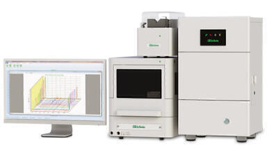
Preparative imaged Capillary Isoelectric Focusing (iCIEF) was performed using a CEInfinite multi-functional system equipped with an energy-efficient LED UV (280 nm) light source and a CMOS camera.
The anolyte was 80 mM phosphoric acid and the catholyte was 100 mM sodium hydroxide. Both solutions were prepared in 0.1% methyl cellulose. The autosampler sample storage temperature was kept at 10°C.
Separation capillaries for iCIEF
WCID cartridge, 100 µm ID, fluoro carbon (FC) coated capillaries is used for all analytical iCIEF. 200 µm ID acrylamide coated (AD) capillary cartridges (AES, cat. no. CP00303) and microtee integrated (AES, cat. no. CP00303M) are used for iCIEF-MS; 320 µm ID AD coated cartridges (AES, cat. no. CP00307) are used for preparative iCIEF. All these WCID cartridges have a 5 cm long separation capillary, and 50 µm ID transfer capillary are assembled for both iCIEF-MS and preparative iCIEF cartridges. The 200 μm AD coated iCIEF-MS cartridge used for iCIEF-MS includes a quartz union (works as a microtee), connecting the make-up solution and transfer capillary to ESI of MS. Both the make-up solution capillary and transfer capillary have a 100μm ID.
iCIEF-MS
A Thermo Exactive Plus EMR mass spectrometer equipped with an Ion Max ESI Ion Source with a 34-gauge needle (Thermo Fisher Scientific, Bremen, Germany) was used for mass measurement. The spray voltage: 3.8 kV, sheath gas: 20 psi, Auxiliary gas: 0, Probe heater: 80°C, Capillary temp: 320°C.
The focusing was 1 min at 1000 V, 1 min at 2000 V and 7 min at 3000 V, and 3000 V during mobilisation; the mobilisation speed was 0.05 µL/min across the separation capillary, and 5 µL/min make up solution added through a micro tee. Mobilisation time was 15 min.
Software
The control and data acquisition of both analytical and prep is with CEInsight, which is CFR21 part 11 compliant. Charge profile data was exported from CEInsight and processed with Clarity (DataApex, Prague, The Czech Republic).
Principle of Preparative iCIEF
Capillary isoelectric focusing is a two-step process. After introducing the sample, 22 µL of premixed solution of the mAb, ampholytes and optional additives, into the separation capillary, the voltage is applied. The sample solution is focused for 1.0 minutes at 1.5 kV and then for 7.5 minutes at 3.0 kV. With the CEInfinite instrument, the focusing process can be monitored live in 15-second intervals. The focusing process is illustrated in the top graphic of Figure 2.
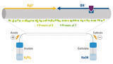
The AES patented capillary diameter transformation technique (CDTT) [4] allows fractionation of protein charge variant purely based on pI. Unlike capillary isoelectric focusing on a CE-instrument, which requires that the catholyte vial is exchanged with a low pH buffer to move the focused zones past the point of detection, in CEInfinite a precise syringe pump is used (indicated in Figure 2), which starts after completion of focusing stage. Mobilisation is performed at 0.16 µL/min for 30 min.
The charge variant peaks are pushed by the syringe pump from the inlet to the outlet of the separation column while maintaining the high voltage. By the continuous migration of hydroxide ions from the cathodic side and protons from the anodic side of the separation capillary, the charge variant peaks in the separation capillary remain focused under the electric field.
The inner diameter of the transfer capillary is much smaller than the separation column. Once exiting from the separation capillary, the charge variant zone length expands by the inner diameter ratio square. For a prep cartridge with a separation capillary of 320 µm ID and a transfer capillary 50 µm, about 41 times peak expansion is expected. The peak expansion inside the transfer capillary, where no electric field is applied, effectively minimises unwanted remixing charge variant zones.
Automated iCIEF fractionation
Unattended automated sample analysis is highly desirable in protein separation and characterisation nowadays. Thanks to the CDTT, the charge variant peaks can be fractioned with high purity. Higher purity charge variant collections can be accomplished by heart cut fractionation. To facilitate collections and to improve fraction collection efficiency, automated fraction collection (AFC) with an integrated selection valve and micro tee (Figure 3a) was developed. A make-up liquid at 10-20x mobilisation flow rate was applied to minimise evaporation of the collected zone. The transfer time from the separation capillary into the collection for charge variant peak is about 2-3 minutes, which allows the completion of charge variant fraction collection within 30 mins.
In Figure 3b the online coupling of iCIEF with ESI-MS is illustrated. A sheath solvent is added in short distance before the mass spectrometer entrance to optimise the conditions for electrospray ionisation.
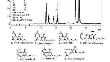
To demonstrate the separation resolution and repeatability of NISTmAb iCIEF, an injection of this sample was repeated three times using a i.d 100 µm FC cartridge. The iCIEF profiles of the three runs are overlaid in Figure 4 to show the reproducibility of the iCIEF analysis of NISTmAb. The five dominant charge variants of mAb A are labeled in Figure 4: the first and second acidic peaks, acidic 1 and acidic 2, respectively; the main peak; and the major and minor basic peaks.
Mobilisation
The mobilisation speed has a significant impact on the efficiency and resolution of prep iCIEF, an elevated pressure and a mobilisation speed which is too high will cause disturbance of the focused protein iCIEF profile, while too low a pressure will reduce the efficiency of the prep iCIEF. The mobilisation speed was optimised and controlled so that overall resolution of protein was preserved.
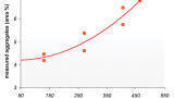
To confirm the charge variants’ purity, the collected fractions were subjected to iCIEF analysis. 5 µL of the collected solution was mixed with 20 µL IEF solution with two pI markers. The top trace in Figure 5 shows the electropherogram of NISTmAb in the prep cartridge during the mobilisation step. The stack of iCIEF profiles for all fractions is shown in the lower trace. Table 2 lists the calculated purity of every variant.
iCIEF-MS
The AES patented CDTT allows a flexible and straightforward iCIEF-MS separation and characterisation of protein charge variant purely based on pI. There are many unparalleled advantages associated with CDTT iCIEF-MS
1. Minimal iCIEF-MS development is necessary. Identical iCIEF conditions can be directly transferred by selecting make up liquid and MS conditions.
2. Protein charge variants were separated in CDTT iCIEF-MS based on pI at iCIEF and measured the corresponding molecular weight at MS. Unlike other CIEF-MS or iCIEF-MS that involves chemical mobilisation, charge variant peaks are sequentially forced by hydrodynamic force from the separation capillary to the transfer capillary. While in the separation capillary, due to the high electric field and continuous migration of hydroxide ions from the cathodic and protons from the anodic side, the charge variant peaks enter the transfer capillary purely based on pI. There is no need for extra electrolyte switching after focusing, since no chemical mobilisation is involved in the mobilisation process. These greatly simplified iCIEF-MS procedures enable straightforward and robust operations.
3. Coupling CDTT iCIEF to MS works more like HPLC-MS than CE-MS. The electric circuit is completed at the separation capillary, and no current is necessary at the transfer capillary. Preferably both ends of the transfer capillary should be grounded. Therefore, there is great flexibility being able to connect the transfer capillary directly to a low flow ESI or nano ESI.
4. It is possible to conduct intact protein iCIEF-MS under denatured (at extreme pH) or native (at neutral pH) conditions with CDTT iCIEF. With direct iCIEF to low flow ESI or nano ESI, a make-up liquid at 1-20 times the mobilisation flow-rate can be applied. The composition of the make-up liquid can be any that is suitable to MS analysis. Consequently, protein charge variant iCIEF-MS can be achieved under denatured or native conditions by controlling the pH and composition of the make-up liquid.
5. CDTT iCIEF-MS permits multiple MS scans of a charge variant peak since once the charge variant peaks enters the smaller ID transfer capillary, it expands inside the transfer capillary. Although there is Tylor dispersion associate with the bulk movement, a higher purity in the centre section of the expanded peak can be expected. The significantly expanded peaks allow more MS scans for each individual peak, and the higher variant purity at the centre section of the expanded peak also facilitates charge variant identification.
6. CDTT iCIEF-MS enables novel applications. Professor Neusus’ group demonstrated iCIEF-CZE-MS. Based on the same principle, iCIEF-nanoHPLC-MS and iCIEF-enzyme reactor transfer capillary-MS are possible.
NISTmAb is a well-studied molecule and was selected to illustrate CDTT iCIEF-MS. The analytical iCIEF required 2M urea and 0.35% MC in the sample solution. Since MC and urea are not MS-compatible, 20% formamide was added to replace the urea and MC. The existing iCIEF method was applied to CDTT iCIEF-MS, with the make-up solution of 1% formic acid and 50% acetonitrile. The mobilisation speed was 0.05 µL/min, and the make-up solution was 5 µL/min. It can be calculated that the eluted charge variant and matrix are diluted about 100 times with the MS compatible solution before the charge variant entered the ESI source. This greatly reduces the risk of ESI source contamination with components in the matrix and effectively prevented potential capillary clogging.
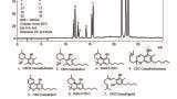
Clean iCIEF-MS signals for all the charge variants were obtained. The top diagram in Figure 6 displayed the iCIEF profile and TIC profile of the iCIEF-MS of NISTmAb. Deconvolution of each charge variant mass spectrum generated multiple masses, corresponding to neutral glycosylation variants’ expected presence. The averaged and deconvoluted mass spectra of each charge variant peak from the iCIEF-MS analysis of intact NISTmAb sample are shown in Figure 7. Overall, the deconvoluted mass spectra’s peak shapes show good peak symmetry, indicating minimal interference from the carrier ampholytes adducts. The identity of each charge variant species was based on the known molecular weight of the main peak and Δmass relative to it. The two basic peaks, basic 1 and basic 2, can be readily assigned as one unprocessed Lys residue at the heavy chain C-terminus and two unprocessed Lys residues at the heavy chain C-terminus, respectively. The deconvoluted mass spectrum of acidic peak 1 showed inconsistent Δmass to that of the main peak. Different Post translational modifications (PTMs) such as deamidation, glycation, cysteinylation, glutathionylation might be expected. Unambiguous identification of these PTMs can be done with other techniques such as iCIEF fraction collection, controlled enzyme digestion, deglycosylation.
Streamlining iCIEF-MS of CDTT iCIEF and Thermo Fisher Scientific’s powerful Orbitrap MS greatly facilitates protein charge variant characterisation. Unattended iCIEF-MS has been enabled with contact closure between the CEInfinite CE system and QEplus MS, providing high resolution and high-efficiency iCIEF-MS for routine characterisation of biopharmaceuticals and other macromolecules. Case studies from universities and biopharmaceutical companies have been presented in recent webinars and conferences to illustrate the strength and application of iCIEF coupled to MS for mapping the main degradation pathways and critical quality attributes of biopharmaceuticals [13,14].
Highly efficient and robust iCIEF have been utilised in biopharma for routine quality and quantify protein charge heterogeneity at any point in biotherapeutic discovery, development, and production. The development and commercialisation of CDTT iCIEF technology described and demonstrated that automatic charge variant fraction collection based on iCIEF profile can be achieved with prep iCIEF in 40 min. The purity of the collected protein variant can be confirmed and used for further intact MS, peptide mapping, and SPR characterisation. The developed iCIEF-MS has been introduced. It has been demonstrated that a highly effective iCIEF-MS of protein charge variant can be realised in 15 min with conventional low flow ESI MS.
1. G. Rozing. Chromatograpy Today. (2019), 8-14.
2. Z. Sosic, D. Houde, A. Blum, T. Carlage, and Y. Lyubarskaya. Electrophoresis. 29 (2008), 4368-76.
3. D. A. Michels, et al. BioProcess International. 9 (2011), 48-54.
4. T. Huang. US Patent # 10935519.
5. R. S. Rogers et al. The AAPS Journal. 20 (2018), 7.
6. C. Montealegre, C. Neusüß. Electrophoresis 39(2018), 1151-1154.
7. Y. Yan, A.P. Liu, S.Wang, T.J. Daly, and N. Li. Anal. Chem. 90(2018), 13013-13020.
8. J.Dai, J. Lamp, Q. Xia, Y. Zhang. Anal. Chem. 90(2018), 2246–2254.
9. J. Dai, Y. Zhang, Anal. Chem. 90(2018),14527-14534.
10. L.Wang, and D.D.Y. Chen. 40(2019), ELECTROPHORESIS. 2899-2907.
11. S. Mack et al. ELECTROPHORESIS. 40(2019), 3084-3091.
.12 T. Huang, XZ. Wu, J. Pawliszyn, Anal. Chem. 72(2000), 4758-4761.
13. C. Neusüß, C. Sönksen, T. Huang. https://www.selectscience.net/expert-insight/the-future-of-cief-mass-spec-techniques-for-protein-characterization/?artID=49878.
14. D.B. Kristensen. Presented at Bioprocessing Summit Europe 2021.