Preparative
Published over 12 years ago. See the latest and most current information on Preparative.
The development of a complete analytical method includes a number of steps from sample collection to the final reporting of results. Intermediate stages involve sample storage, sample preparation, isolation of analytes, their identification and quantification. However, sample preparation is one of the most crucial processes in bioanalytical applications. The results of the experiment depend on the condition of the starting material, thus, a proper experimental model and careful sample preparation is crucial in order to obtain significant and reliable results. In the context of protein science and proteomics research, phosphoproteomics has become an important research area in several life sciences. However, phosphorylated compounds are usually present at very low concentrations. Thus, highly sensitive and selective technologies are required for their enrichment and purification. This review provides a short overview about new developments in sample preparation for the enrichment of phosphorylated peptides and proteins.
Biological samples used as a source for bioanalytical applications are usually very complex and need a prior clean-up or enrichment procedure in order to reduce sample complexity. There is a need for highly efficient bioanalytical approaches to handle the problems associated with sample preparation, separation and identification of proteins and peptides within biological species. Many newly emerged technologies meet the basic requirements and considerable progress has been made in the development of new stationary phases for the enrichment of phosphorylated peptides and proteins. Although many biochemical mechanisms are involved in cellular signalling, reversible phosphorylation of serine, threonine, and tyrosine residues is one of the most commonly used in mammalian cells (Figure 1) [1, 2]. The analysis of the entire cellular phosphoproteins panel, the so-called phosphoproteome, has been an attractive study subject since the discovery of phosphorylation as a key regulatory mechanism of cell life [3, 4, 5]. The high importance of protein phosphorylation makes the comprehensive identification of phosphorylation sites an important task for understanding biological functions. The covalently attached phosphorylation sites are usually present at substoichiometric levels. This means that a post-translational modification at a given site is often present in only a small fraction of the protein molecules of a given type. Thus, the development of sensitive and selective technologies which allows the enrichment of phosphorylated proteins or peptides is of utmost importance [6].
New enrichment tools in phosphoproteomics
As a consequence, novel enrichment and desalting methods based on modern solid-phase extraction (SPE) technologies have been designed for the purification of biological samples which can be tailored to a specific bioanalytical application allowing endless possibilities in terms of selectivity tuning [7, 8]. SPE is a fast, versatile, easy to use and easy to automate sample preparation technique and enables both, concentration and purification of the sample. In particular, nanomaterials may have a great impact on future sample preparation due to their unique physical and chemical characteristics. Moreover, the move towards miniaturisation in bioanalysis has prompted the development of advanced enrichment and separation technologies by significantly enhancing the hyphenation to mass spectrometry. However, very often buffered solutions, salts and detergents are needed during the sample preparation procedure which tend to interfere with further protein identification, as they might inhibit the digestion process or disturb the mass spectrometric analysis. Usually a combined desalting and concentration step is performed in order to remove potentially interfering detergents and salts. This can be achieved by dialysis [9, 10], diafiltration [11, 12], gel filtration [13, 14] and centrifuge columns
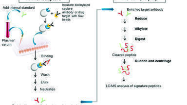
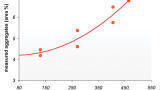
[15, 16]. Alternative chromatographic materials for desalting include reversed phase chromatography [17, 18], oxidised diamond particles [19], magnetic beads [20, 21] and C60-fullerene silica [22]. For the identification of phosphoproteins mass spectrometry has become the most powerful technique [23]. However, mass spectrometric detection of phosphorylated peptides in complex samples represents a major challenge due to their low abundance in comparison to other non-phosphorylated peptides. Very often the signals of phosphorylated peptides are suppressed by the signals of higher-concentrated non-phosphorylated peptides [24]. A major strategy to overcome this problem includes the application of selective enrichment technologies for phosphorylated proteins and peptides before MS analysis. A number of different techniques have been developed to enrich phosphoproteins or phosphopeptides from biological samples. These include antibody-based immunoaffinity precipitation [25], chemical modification [26], enrichment on immobilised metal ion affinity chromatography (IMAC) [27, 28, 29], enrichment on metal oxides [30, 31, 32], or coupling IMAC and metal oxide enrichment [33]. SPE has been performed with various nanomaterials, embedded or coated on the walls of monolithic tips. Pipette tips were fitted with a monolithic structure based on poly(divinylbenzene) containing embedded TiO2 and ZrO2 nanoparticles [34]. The micropipette tip format permits the handling of submicrolitre amounts of samples and offers the suitability for further combination with automated robotic systems, which leads to higher reproducibility and shorter time of sample preparation. The effect of particle size on the selectivity of phosphopeptides was investigated by comparative studies with nano- and microscale TiO2 and ZrO2 powders. In this study nanomaterials performed much better in terms of binding capacity. When compared to conventional Fe-IMAC, higher selectivity was observed in case of monolithic TiO2/ZrO2 tips. Recently, a new type of IMAC adsorbent using Zr4+ and Ti4+ ions was developed on the basis of phosphonate groups [35, 36]. In particular Ti4+-IMAC showed superior performance compared to conventional Fe3+-IMAC and TiO2. In a further approach trivalent lanthanum, holmium and erbium ions were chelated to a highly porous phosphonate polymer which was prepared by radical polymerisation of vinylphosphonic acid (VPA) and divinylbenzene (DVB) [37]. Lanthanides are known to be hard “acceptors” with an overwhelming preference for oxygen-containing anions such as phosphates. A major advantage of lanthanide ions for IMAC is their high coordination number, resulting in stronger interactions with phosphopeptides. Lanthanides offer six to twelve coordination sites with eight and nine being the most common coordination numbers [38, 39]. Due to their increased number of coordination sites they significantly enhance the binding of phosphorylated peptides. Compared to commonly used TiO2, the lanthanide-IMAC strategy showed higher selectivity for phosphorylated peptides. In another study, lanthanum-functionalised diamond-IMAC was successfully applied for trapping phosphopeptides from bovine milk and egg-yolk [40]. Moreover, lanthanum silicate based magnetic affinity microspheres could be successfully applied for isolating phosphopeptides from complex protein digests [41]. Despite recent advances in phosphopeptide research, detection and characterisation of multiply phosphorylated peptides have been a major challenge. A number of strategies have been developed for detecting multiply phosphorylated peptides. For example, phosphoric acid was used as a matrix additive for improving the MALDI signals of phosphopeptides in the presence of non-phosphorylated peptides [42]. In another approach derivatised polymer brushes with oxotitanium and nitrilotriacetate-Fe(III) groups were found to enrich both, mono- and multiply phosphorylated peptides [43]. Recently, C60-fullerene-aminopropylsilica was shown to have a preferential enrichment potential for multiply phosphorylated peptides which are usually difficult to detect. The fractionation of mono- from multiply phosphorylated peptides occurs due to the different pI values of the peptides and the effect of the total charge of the peptides. Multiply phosphorylated peptides usually have a low pI (the isoelectric point - the pH at which the molecule carries no net electrical charge) value and basic elution (e.g. ammonia solution, pH 10) mainly retrieves peptides with a pI < 3. A second elution step was achieved at pH 2.3 using 1% TFA containing 20% ACN and resulted into the release of monophosphorylated peptides. Finally, non-phosphorylated peptides could be eluted in 80% ACN/1% TFA. Figure 2 depicts commonly applied tools for phosphopeptide purification as well as innovative enrichment strategies such as La-IMAC and C60-fullerene silica.
Separation of enriched molecules
Many bioanalytical applications still require a further separation of enriched molecules particularly for their identification, structural elucidation and quantification. For that novel polymeric capillary monoliths which are tailored for miniaturised liquid chromatography (μ–LC) offer a highly efficient unique separation tool [44]. The influence of different polymerisation parameters like monomer to porogen ratio, microporogen type, microporogen content and polymerisation temperature on the porous structure enabled systematic optimisation of the separation performance. In this regard, the significant reduction in polymerisation time was observed resulting in the formation of a high fraction of small macropores while, at the same time, keeping the fraction of flow-channels reasonably high [45, 46]. Separation efficiencies of monolithic capillaries are outstanding as they have no inter-particular void volume and retaining frits, which results in a reduced resistance to mass transfer as a result of convective flow.
References
[1] T. Hunter, Cell 100 (2000) 113-127.
[2] M.J. Hubbard, P. Cohen, Trends Biochem. Sci. 18 (1993) 172-177.
[3] S.A. Johnson, T. Hunter. Nat. Methods 2 (2004) 17-25.
[4] G. Manning, D.B. Whyte, R. Martinez, T. Hunter, S. Sudarsanam, Science 298 (2002) 1912-1934.
[5] T. Pawson, N. Piers, Genes Dev. 14 (2000) 1027-1047.
[6] M. Mann, S.E. Ong, M. Grønborg, H. Steen, O.N. Jensen, A. Pandey, Trends Biotechnol. 20 (2002) 261-268.
[7] S.H. Bae, A.G. Harris, P.G. Hains, H. Chen, D.E. Garfin, S.L. Hazell et al., Proteomics 3 (2003) 569-579.
[8] T. Ba̧czek, J. Pharm. Biomed. Anal. 34 (2004) 851-860.
[9] H. Görisch, Anal. Biochem. 173 (1988) 393-398.
[10] W. Qinyuan, C. Liu, R.D. Smith, Rapid Commun. Mass Spectrom. 10 (1996) 835-838.
[11] A. Simon, L. Vandanjon, G. Levesque, P. Bourseau, Desalination 144 (2002) 313-318.
[12] L. Schwartz, Pall Scientific & Technical Report (2003).
[13] J. Porath, P. Flodin, Nature 183 (1959) 1657-1659.
[14] G. Bertil. J. Chromatogr. A 3 (1960) 330-342.
[15] R.I. Christopherson, M.E. Jones, R.F. Lloyd, Anal. Biochem. 100 (1979) 184-187.
[16] E. Helmerhorst, G.B. Stokes, Anal. Biochem. 104 (1980) 130-135.
[17] T. Pohl, R.M. Kamp. Analytical Biochem. 160 (1987) 388-391.
[18] M. Palmblad, J.S. Vogel, J. Chromatogr. B 814 (2005) 309-313.
[19] X.L. Kong, L.C.L. Huang, C.M. Hsu, W.H. Chen, C.C. Han, H.C. Chang, Anal. Chem. 77 (2005) 259-265.
[20] H. Chen, C. Deng, X. Zhang, Angew. Chem. Int. Ed. 49 (2010) 607-611.
[21] W.Y. Chen, Y.C. Chen, Anal. Chem. 79 (2007) 8061-8066.
[22] R.M. Vallant, Z. Szabo, S. Bachmann, R. Bakry, M. Najam-ul-Haq, M. Rainer et al. Anal. Chem. 79 (2007) 8144-8153.
[23] J.D. Dunn, G.E. Reid, M.L. Bruening, Mass Spectrom. Rev. 29 (2010) 29-54.
[24] D.T. McLachlin, B.T. Chait, Curr. Opin. Chem. Biol. 5 (2001) 591-602.
[25] Z. Guoan, T.A. Neubert, Proteomics 6 (2006) 571-578.
[26] O. Yoshiya, T. Nagasu, B.T. Chait, Nat. Biotechnol. 19 (2001) 379-382.
[27] J. Porath, J. Carlsson, I. Olsson, G. Belfrage, Nature 258 (1975) 598-599.
[28] M.C. Posewitz, P. Tempst, Anal. Chem. 71 (1999) 2883-2892.
[29] I. Feuerstein, S. Morandell, G. Stecher, C.W. Huck, T. Stasyk, H.L. Huang et al., Proteomics 5 (2005) 46-54.
[30] A. Leitner, Trends Anal. Chem. 29 (2010) 177-185.
[31] M.R. Larsen, T.E. Thingholm, O.N. Jensen, P. Roepstorff, T.J.D. Jørgensen, Mol. Cell. Proteomics 4 (2005) 873-886.
[32] H.K. Kweon, K. Hkansson, Anal. Chem. 78 (2006) 1743-1749.
[33] T.E. Thingholm, O.N. Jensen, P.J. Robinson, M.R. Larsen, Mol. Cell. Proteomics 7 (2008) 661-671.
[34] M. Rainer, H. Sonderegger, R. Bakry, C.W. Huck, S. Morandell, L.A. Huber, D.T. Gjerde and G.K. Bonn, Proteomics 8 (2008) 4593-4602.
[35] H. Zhou, S. Xu, M. Ye, S. Feng, C. Pan, X. Jiang et al. J. Proteome Res. 5 (2006) 2431-2437.
[36] J. Dong, H. Zhou, R. Wu, M. Ye, H. Zou, J. Sep. Sci. 30 (2007) 2917-2923.
[37] M.R. Mirza, M. Rainer, C.B. Messner, Y. Güzel, D. Schemeth, T. Stasyk et al., Analyst 138 (2013) 2995-3004.
[38] Z. Ahmed, K. Iftikhar, Inorg. Chem. Commun. 13 (2010) 1253–1258.
[39] F.S. Richardson, Chem. Rev. 82 (1982) 541-552.
[40] D. Hussain, M. Najam-ul-Haq, F. Jabeen, M.N. Ashiq, M. Athar, M. Rainer et al., Anal. Chim. Acta 775 (2013) 75-84.
[41] G. Cheng, Y.L. Liu, J.L. Zhang, D.H Sun, J.Z. Ni, Anal. Bioanal. Chem. 404 (2012) 763-770.
[42] S. Kjellström, O.N. Jensen, Anal. Chem. 76 (2004) 5109-5117.
[43] W. Wang, J. Dong, G.L. Baker, M.L. Bruening, Analyst 136 (2011) 3595-3598.
[44] L. Trojer, S.H. Lubbad, C.P. Bisjak, W. Wieder, G.K. Bonn, J. Chromatogr. A 1146 (2007) 216-224.
[45] L. Trojer, C.P. Bisjak, W. Wieder, G.K. Bonn, J. Chromatogr. A 1216 (2009) 6303-6309.
[46] A. Greiderer, S.C. Ligon, C.W. Huck, G.K. Bonn, J. Sep. Sci. 32 (2009) 2510-2520.