Electrophoretic separations
Published over 15 years ago. See the latest and most current information on Electrophoretic separations.
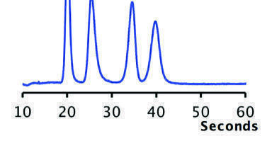
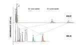
This article shows how a capillary electrophoresis instrument may be used in conjunction with UV area imaging to determine mobility, diffusion coefficient and charge of components in a mixture of small molecules. The analytes lidocaine, phenylmethanol and benzoate are chosen as representative of small molecules of different charge type: cationic, neutral and anionic respectively in the background electrolyte used, phosphate buffer at pH 7.5. A capillary with three windows is used, with detection at the first window using the Agilent 7100 CE system and at the second and third using the ActiPix D100 UV imaging detector. The separated species are characterised at the first window after short-end injection followed by pressure assisted capillary electrophoresis for 3.5 minutes. The diffusion coefficients and hydrodynamic radii of the molecules are determined from the broadening of the bands between the second and third windows during a 15 minute stage of pressure driven flow. The method allows diffusion coefficients of all species to be measured within a single run, with good repeatability (RSDs better than 2.5%, n=9). Mobilities are found from times to these windows in separate experiments using pressure assisted capillary electrophoresis. Charge follows by combining data on diffusion coefficient and mobility.
Introduction
There is a need within the biopharmaceutical and pharmaceutical industries for techniques which allow diffusion coefficients and electrophoretic mobilities of molecules to be determined in a single experimental system.
Knowing the ratio of mobility to diffusion coefficient allows the charge, or valence, of the molecule to be determined [1]. For antibodies, the higher the valence, the less the tendency for aggregation [2].
Capillary electrophoresis (CE) is a well established technique for separating compounds by application of voltage, and the method is used to measure electrophoretic mobilities with high precision [3,4]. CE instruments also allow application of pressure, and analysis of band broadening due to Taylor dispersion during pressure-driven flow in a standard CE setup provides data on diffusion coefficient and hydrodynamic radius [5-10].
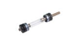
There has been one report on use of a CE system with a mixture of components, using initial application of voltage to separate them followed by application of pressure to broaden the bands and allow determination of diffusion coefficients by Taylor dispersion analysis (TDA) [9]. The aim of this article is to extend this approach, using the pairing of Paraytec’s ActiPix D100 UV imaging detector and the Agilent 7100 CE system. This CE system has been designed to allow use with external detectors. In previous work we have shown that use of the ActiPix UV imaging detector with two windows in a single capillary allows hydrodynamic radii to be determined with high precision in a single experiment [11,12]. Our objective in this article is to illustrate the approach and methodology for measurement of charges of individual components in a mixture of small molecules.


-(1).jpg)

2.jpg)