Data handling
Published over 4 years ago. See the latest and most current information on Data handling.
One of the most important assays in the development of any biotherapeutic protein is peptide mapping. The analysis is used to confirm that the correct protein sequence has been expressed and provides structural characterisation of amino acids and post-translational modifications (PTMs). Advances in mass spectrometry (MS) and separation technologies are enabling the generation of comprehensive peptide maps for confident biotherapeutic quality control (QC)/assurance. High-Resolution Ion Mobility (HRIM) coupled with MS provides improved/more accurate peak identification and quantitation compared with traditional liquid chromatography (LC) methods. This article will explain why the use of a simple, easy-to-implement HRIM-MS method should be implemented into PTM analysis and show evidence of the benefits over LC-only detection.
Introduction
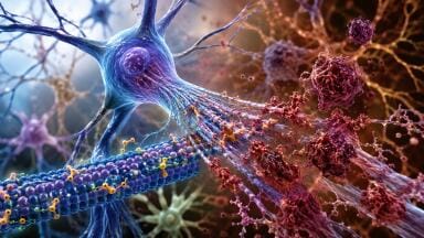
Biotherapeutic medicines, like monoclonal antibody (mAb) products, biosimilars and cell and gene therapies, are of increasing interest due to their ability to target specific molecules within the human body and their good track record with patient safety [1]. The quality of these medicines is vital to ensure their safety and efficiency. Most proteins used in medicines adopt a specific three-dimensional structure under physiological conditions. Post-translational modifications (PTMs) to amino acid side chains caused by external environmental factors, such as excessive heat or light, can disrupt the secondary and tertiary structure of the protein leading to decreased efficacy or safety of a protein- or peptide-based therapeutic product [2]. For this reason, federal health authorities require demonstration of complete product knowledge, including correlation of forced degradation to antigen binding, at the time of filing.
Peptide mapping is used to determine the fingerprint of the protein, which confirms its primary structure and detects alterations in primary structure. It is important to know the structure of the protein to determine if or how it has changed during production, processing or storage, all of which may affect the function of the protein. Peptide mapping and PTM analysis also allow for monitoring critical quality attributes (CQAs) and understanding the drug product and its structure, stability, efficacy, and function.
This is extremely important in drug products, not only in the research and development stage, but also in the commercialisation, manufacturing, and QC space so that products can be scaled up accurately. In QC, identity is confirmed when the chromatographic profile of a peptide map conforms to expectation in comparison to a reference map (e.g., peak retention time, peak height, no new or missing peaks) [3]. CQAs are being monitored during a product’s lifecycle to ensure its consistency and quality.
Peptide mapping is carried out as shown in Figure 1. A therapeutic protein is digested into constituent peptides using chemical or enzyme reactions. Once digested, the solution contains thousands of peptides that require separation, so a reliable separation platform that can deliver the highest possible resolution is needed – this is usually high-performance liquid chromatography (HPLC). The sample is then injected into a mass spectrometer instrument, such as a time-of-flight (ToF), where different peptides can be detected and analysed with high mass accuracy. Data processing strategies are then employed to analyse the data from the instrumentation. The greater the number of matches between the experimental and database peptide masses, the higher the confidence in the protein’s identification.
Complex workflows require higher resolution separations
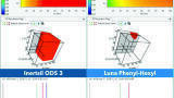
LC-MS provides the potential for rapid, cost-effective, and quantitative measurements of organic molecules for an enormous variety of applications. As a result, it has become the ‘go-to’ technique for many quantitative analysis applications. Whilst there is no doubt that modern LC-MS instruments are relatively easy to operate and maintain, there are also many difficulties, especially for accurate quantitative measurement of analytes in complex samples. Peptide mapping requires shallow analytical gradients, which can take, on average, upwards of 90 minutes per sample to resolve near isobaric PTMs. Examples of this are asparagine deamidation, or isobaric PTMs, such as aspartate isomerisation. The level of detail required comes at the expense of analysis time. Even with long analysis times, some deamidation and isomerisation events go undetected due to lack of chromatographic resolution. When developing a peptide mapping method, the use of sample preparation conditions that will minimise the degree of artificial modifications induced is critical. LC often necessitates matching column size, length, and packing material to specific separations and their intended purposes - all of which have the potential to introduce complexities into the workflow.
A novel approach
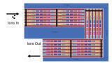
Unlike traditional reversed-phase LC, which separates molecules based on hydrophobicity in the condensed-phase, Structures for Lossless Ion Manipulation (SLIM)-based HRIM-MS separations sort molecules by their size, shape, and charge post-ionisation, making it an ideal, orthogonal complement to LC methods. Like other ion mobility (IM) techniques, ions are propelled through an inert buffer gas, such as nitrogen or helium, and along a flight path by the application of an electric field. The ions experience collisions with the buffer gas, with small ions traversing the path faster than large ions. Ions will experience more collisions over longer IM path lengths, thus increasing peak resolving power. However, with existing IM technologies, there are concerns about ion loss leading to a decrease in sensitivity, the mass range per analysis is limited, and to attain high-resolution separation, the length of the ion path must be exceptionally long, which logistically is not possible.
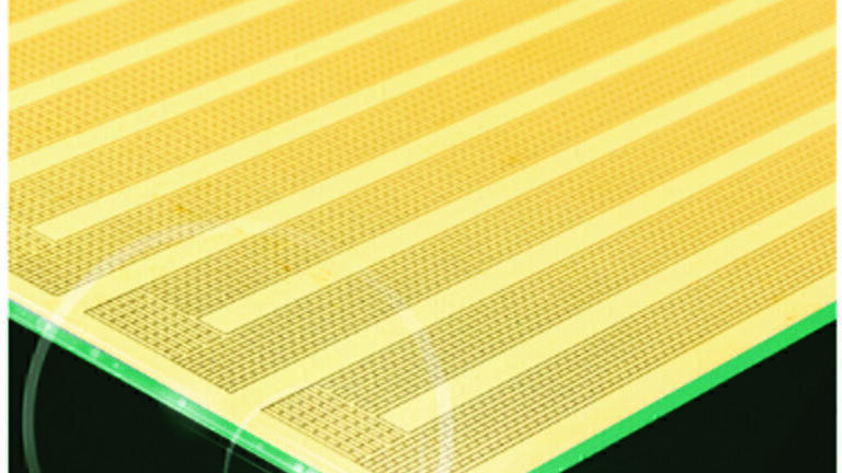
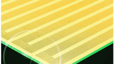
HRIM-MS provides an exceptionally long ion path (13 meters) delivering superior IM resolution and the ability to rapidly and reproducibly separate and identify isobaric PTMs. The system uses electrode patterns on standard printed circuit board (PCB) technology (Figure 2). Digitising separations in this way on PCBs addresses the ion loss and resolution limitations of other IM technologies, resulting in superior separation of challenging molecules of interest. The electric fields that propel the ions also prevent them from striking surfaces while moving, therefore preventing any losses along the way. The unique serpentine path design of the PCBs permits a 13m ion path, allowing for unparalleled separation of isomeric structures.
The key advantage of implementing HRIM in the peptide mapping workflow is that lengthy chromatographic gradients are no longer required to resolve isobaric PTMs; these are resolved in the IM dimension instead, so analytical gradients can be shortened without loss of peak separation. This reduced reliance on LC separations helps ease potential bottlenecks in analytical workflows.
HRIM allows analytical run times of minutes compared to hours, dramatically increasing throughput. For example, a typical 60–90-minute peptide map is accomplished in five minutes with greater PTM elucidation, which is 2-3x faster than a traditional LC experiment. It provides a significant increase in throughput, enabling more samples to be analysed in a more efficient manner. In the fast-paced area of biotherapeutic development, this enables the screening of more drug candidates or analysing more patient samples in a shorter time.
The incorporation of HRIM into the workflow allows for reduced reliance on LC for the separation of complex biological matrices. This greatly improves reliability in the lab and also provides superior reproducibility. With better reproducibility comes more confidence in results, which is especially crucial in the QC space, where high resolution data needs to be obtained with the least amount of noise.
Here we describe how a HRIM-MS platform can be used as a high throughput alternative to long LC-MS gradients by providing a peptide digest mass analysis protocol.
HRIM-MS in practice
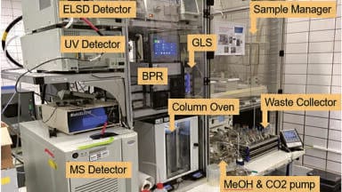
LC-HRIM-MS methods were performed on a HRIM instrument (MOBIE, MOBILion Systems Inc, Chadds Ford, PA) coupled to an Agilent 6545XT Q-ToF (Agilent Technologies, Santa Clara, CA). A schematic of the HRIM instrument connected to an Agilent 6545XT Q-ToF is shown in Figure 3. The HRIM instrument nests between the Agilent ion source and the Q-ToF ion optics. Reversed-phase chromatography was performed using a C18 column 2.1 x 50mm, 1.8 μm Zorbax® Extend C18 column (Agilent Technologies) with a 20-minute gradient. The data was recorded in a proprietary data format and converted to the Agilent IM-MS MassHunter data format (.d). Sequence coverage was determined using Skyline software. Peptide PTM arrival times were extracted by m/z from MassHunter IM-MS browser software (Agilent Technologies) [4].
Figure 4 shows a full chromatogram where different peaks for each peptide are coming off the instrument, with the MS data and mobility data underneath. On closer inspection of the data shown between 23 and 27 minutes, highlighted by the blue box, and summed up by the MS data, the individual peptides are visible in good resolution, with baseline resolved peaks. However, the peptides that have been identified are showing a 1 Da mass shift, which is indicative of deamidation.
Examining the data at 30.5–36 minutes (Figure 5) on specific peptides and the 1 Da shift, the mobility data shows the unmodified and deamidated versions as well as a deamidated version with a sodium adduct. The HRIM instrument is giving the ability to identify deamidation, without needing to run complex instrumentation such as Fourier-transform ion cyclotron resonance or ion trap mass analysers. In chemistry, manufacturing, and control (CMC) where reproducibility and ease of use is needed, it is necessary to have the ability to be able to detect with confidence and accuracy. The high resolution, high availability data that comes from HRIM, as well as the ability to look at the mobility data, helps identify putative deamidation domains without complex instruments and the operators who are needed to run them. The data provided not only gives high resolution for the mass of the molecule, but also information and confirmations of the structure which is important when ‘structure equals function’.
Demonstrating the utility of HRIM
Traditional LC-MS peptide mapping workflows allowing for full sequence coverage and characterisation of PTMs have gradient times of 60–120 minutes per sample [3, 5, 6, 7] and above [3]. Faster 20-minute methods have been published [8] that deliver full sequence coverage, but they do not resolve deamidation and isomerisation of PTMs. Implementing HRIM into this workflow provides an additional dimension of separation, enabling resolution of isobaric species undetected by LC-MS alone in under 20 minutes.
Using the HRIM dimension in combination with the chromatographic dimension allows for dramatically reduced run times without sacrifice in peptide PTM identification or quantitation. Using common LC-MS methods only, the identification of mass-neutral PTMs such as isomerisation are also at risk of being underreported or missed altogether. This clearly demonstrates the utility of HRIM to separate and detect coeluting isomerisation PTMs that are otherwise missed by LC-MS alone.
Peptide mapping and PTM analysis are critical for the monitoring of biotherapeutic quality and their CQAs. Challenges associated with the most common analysis methods include the ability to only analyse the primary structure, long analysis times and difficulty identifying PTMs with similar nominal mass. However, this study has demonstrated that HRIM technology allows for successful analysis that prevents these difficulties. It allows analytical run times of minutes compared to hours and the removal of LC during intact mass analysis, thus preventing issues with LC clogging. Additionally, HRIM also allows for structural information to be obtained on mAbs and for the identification of PTMs such as deamidation.
Getting biopharmaceutical products to market faster
Here we described a mAb tryptic digest, where HRIM enabled critical structural information to be seen that would have been missed using conventional LC-MS techniques alone. Incorporating HRIM into an LC-MS workflow provided a reduction in analysis time from traditional peptide mapping workflows, achieving >96.5% sequence coverage in just 20 minutes total analysis time. HRIM enables rapid PTM identification and monitoring with unparalleled structural resolution. The increased confidence in PTM assay characterisation and quantification enables a more robust approach to biopharmaceutical process monitoring and QC laboratories.
The potential that HRIM holds for the future of biotherapeutics industry is immense. The use of biosimilars, cell and gene therapies and mAbs to treat diseases is a growing trend, and HRIM is transforming the analysis process. Streamlining MS methods for biopharmaceutical analysis is critical to provide rapid, high-quality results to provide safer, more efficacious therapeutic products to market faster.
For more information about HRIM, please visit https://www.mobilionsystems.com/.
References
1. D. E. Johnson, ‘Biotherapeutics: Challenges and Opportunities for Predictive Toxicology of Monoclonal Antibodies’, Int. J. Mol. Sci., vol. 19, no. 11, Nov. 2018.
2. G. A. Khoury, R. C. Baliban, and C. A. Floudas, ‘Proteome-wide post-translational modification statistics: frequency analysis and curation of the swiss-prot database’, Sci. Reports 2011 11, vol. 1, no. 1, pp. 1–5, Sep. 2011.
3. T. Mouchahoir and J. E. Schiel, “Development of an LC-MS/MS peptide mapping protocol for the NISTmAb,” Analytical and Bioanalytical Chemistry, vol. 410, no. 8, p. 2111–2126, 2018.
4. J. Arndt, et al. High-Resolution Ion-Mobility-Enabled Peptide Mapping for High-Throughput Critical Quality Attribute Monitoring. https://doi.org/10.1021/jasms.0c00434.
5. L. W. Dick, D. Mahon, D. Qiu and KC. Cheng, ‘Peptide mapping of therapeutic monoclonal antibodies: improvements for increased speed and fewer artifacts’, J. Chromatogr. B. Analyt. Technol. Biomed. Life Sci., vol. 877, no. 3, pp. 230–236, Jan. 2009.
6. W. Li et al., ‘Structural elucidation of post-translational modifications in monoclonal antibodies’, ACS Symp. Ser., vol. 1201, pp. 119–183, 2015.
7. Q. Dong et al., ‘The NISTmAb tryptic peptide spectral library for monoclonal antibody characterization’, MAbs, vol. 10, no. 3, pp. 354–369, Apr. 2018.
8. D. L. Wong and J. Chen, ‘Making Peptide Mapping Routine with the Agilent 6545XT AdvanceBio LC/Q-TOF Application Note’.


-(1).jpg)