Bioanalytical
Published over 7 years ago. See the latest and most current information on Bioanalytical.
Monoclonal antibodies (mAbs) encompass a rapidly growing therapeutic market. While new antibodies are continually being discovered, expiring patents of the earliest antibodies have prompted the emergence of generic products, or biosimilars. As such, biosimilars are becoming as a key component of major biopharmaceutical companies drug pipelines. Regulatory aspects from the Food and Drug Administration (FDA) [1] and European Medical Agency (EMA) [2] related to the approval of follow on biologics, or biosimilars, specify that the innovator and biosimilar must demonstrate comparability, or similarity, with no clinical meaningful differences. This means that purity, chemical identity, and bioactivity must be characterised. Physicochemical properties are determined by a range of techniques and the data must confirm high similarity to the reference product, or innovator. However, the guidelines are not concrete with respect to analytical technique requirements meaning each company can determine their own set of analytical parameters. Orthogonal analytical techniques are often used in parallel to provide more inclusiveness.
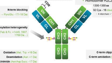
Because mAbs are such large proteins, creating an exact replica is nearly impossible. While the primary amino acid sequence should remain largely the same, post translational modifications (PTMs), like glycosylation, will vary depending on the cell line and manufacturing processes used [3]. These variations make it is necessary to fully characterise new biosimilars in relation to the reference product. The typical physicochemical properties required for protein characterisation, the data includes molecular weight (size), isoform pattern, molar absorptivity, electrophoretic patterns, liquid chromatographic patterns, and spectroscopic profiles. This complex array of data is extensive, but ultimately is used to determine the composition, physical properties, and primary structure of a biosimilar product.
Two critical quality attributes (CQAs) of a biologic that have implications in immunogenicity, safety, and efficacy are protein charge variants and protein aggregates/fragments. These CQAs pertain to the higher-order structure of the protein and are often assessed through chromatographic techniques. Herein, we show charge heterogeneity and aggregation analysis of a therapeutic mAb that recently came off patent, infliximab, as well as two other biosimilars.
Sodium phosphate monobasic, sodium phosphate dibasic, and 2-(N-morpholino)ethanesulfonic acid (MES) hydrate were purchased from Sigma Aldrich (St. Louis, MO). Sodium chloride was purchased from VWR (Radnor, PA). CX-1 pH gradient buffers were purchased from Thermo Fisher Scientific (Waltham, MA). Sodium hydroxide was purchased from Fisher Scientific (Hampton, NH). Infliximab and biosimilars were purchased from Myoderm. A bioZen 6 µm WCX-column (250 × 4.6 mm dimension) was used for all ion-exchange separations (Phenomenex, Torrance, CA). A bioZen 1.8 µm SEC-3. column (300 × 4.6 mm dimension) was used for all size exclusion chromatography (Phenomenex, Torrance, CA). All samples were analysed on an Agilent 1260 HPLC with a chilled autosampler and variable wavelength detector set at 280; data was collected using ChemStation software (Rev. B 04.03-SP1[87], Agilent). The mobile phases for ion-exchange separation chromatography was 20 mM MES pH 5.9 and 20 mM MES 300 mM NaCl pH 5.9. The mobile phase for size exclusion chromatography was 50 mM sodium phosphate pH 6.8 with 300 mM sodium chloride in water running at a flow rate of 0.35 mL/min.
Charge heterogeneity is a critical quality attribute that must be assessed in protein therapeutic development. During the recombinant generation of mAbs, chemical modifications of specific amino acids, e.g. deamidation, methionine oxidation, C-terminal lysine truncation, etc., lead to protein derivatives with slightly different physicochemical properties, namely acidic and basic charge variants. A number of chemical degradation pathways, whether enzymatic or spontaneous, give rise to this heterogeneity and leads to overall variation in isoelectric points (pIs). Two analytical techniques are commonly used for the analysis of charge variants: cation-exchange chromatography (CEX) [4] and imaging capillary electrophoresis (iCE) [5]. While iCE is an excellent technique with high precision, it often lacks robustness, and more importantly does not allow for fractionation and isolation of charge variants. This latter drawback makes the CEX method more utilised in the industry.
Weak cation-exchange (WCX) is the most common chromatographic technique for charge heterogeneity determination. Depending on the stage of development, the chromatographic criteria may differ. For example, during the analytical characterisation stage it is important for precise, reliable, and robust results and therefore longer analysis times are not as critical and are not perceived as being objectionable. During manufacturing however, groups such as process analytical technology require rapid analysis and charge variant results so that production conditions can be altered in real-time. Regardless of the desired outcome, reproducibility and robustness of the method is essential. Two critical aspects of ion-exchange technologies that enable consistency of the technique is particle size distribution and controlled grafting of the stationary phase. For particle size distribution,
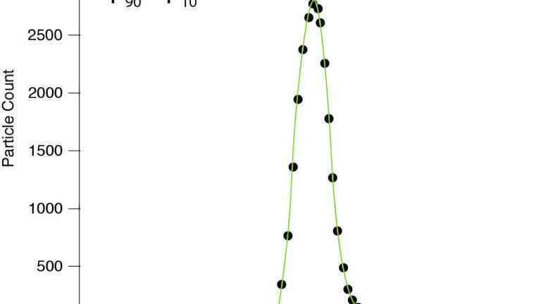
dp90/dp10 analysis is performed on the particle which determines the Gaussian distribution, and thus heterogeneity of the particle size [6]. The closer the dp90/dp10 is to a value of 1.0, the more consistent the particle size. Figure 1 demonstrates a 6 µm non-porous polystyrene divinylbenzene particle the dp90/dp10 of 1.08. This particle combined with a controlled grafting process of the polycarboxylate stationary phase provides a suitable technology for consistent charge variant analysis.
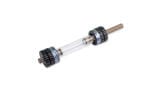
Regardless of the particle technology, one challenge often encountered with charge variant analysis is the creation of an inert column hardware surface. Proteins and their variants tend to interact with trace iron content in column hardware, therefore a bioinert polyether ether ketone (PEEK) column hardware is typically employed. This hardware surface provides excellent recovery of protein charge variants with minimal column priming required. Unfortunately, as laboratories move toward more modern instrumentation, including ultra-high-performance liquid chromatography (UHPLC), to speed up their analyses, PEEK hardware and its back pressure limitations combined with increased column-to-column inner diameter variability can become a problem. A recent development in column hardware uses Medical Grade 2 titanium instead of stainless steel for all column components, including the column tube and frit, which offers similar biocompatibility to PEEK, but with the robustness and pressure capacity of stainless-steel hardware.
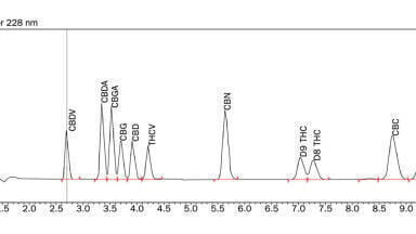
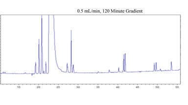
We initiated the charge heterogeneity analysis of infliximab and biosimilars with the 6 µm WCX bioinert titanium column was run at optimal reduced linear velocity of 1 ml/min. Mobile Phase A was 20 mM MES pH 5.9, and Mobile Phase B was 20 mM MES pH 5.9 with 300 mM and a linear gradient was run from 5-35% B in 30 minutes (Figure 2). Under these conditions, the infliximab innovator generated 14% acidic variants and 49% basic variants. Significant differences in charge variants were observed with biosimilars A and B. In both cases, more acidic and fewer basic variants were observed with biosimilar A providing the most acidic variants (31%) and fewest basic variants (9%).
Because mobile phase composition can significantly influence cation-exchange chromatography, we also employed a linear pH gradient to confirm the accuracy of the results. Utilising CX-1 buffers at pH 5.6 and pH 10.2 (Figure 3), running at 1 ml/min and using a linear gradient from 0-100% B we observed comparable results to the salt gradients. The innovator provided identical acidic variants, but fewer basic variants (47%), likely due to differences in C-terminal lysine variants. The biosimilars A and B were also similar to the salt gradients, thus showing that mobile phase choice has minimal influence on the charge heterogeneity results.
The measurement of high molecular weight (HMW) aggregate and low molecular weight (LMW) fragment is performed at many stages in biotherapeutic development. Techniques including sedimentation velocity analytical ultracentrifugation (svAUC) [7] and size exclusion chromatography (SEC) [8] are common methods to assess aggregation. Size exclusion chromatography is a versatile and robust method to determine levels of aggregation and also has the versatility to enable collection of fractions or detection by mass spectroscopy (MS) for more insightful understanding of the material, if desired. Monoclonal antibody therapeutics are primarily immunoglobulin G (IgG) species, which have a molecular weight around 150 kDa. Given the propensity of these mAbs to form dimers, trimers, and tetramers, a SEC particle with the proper pore size to accommodate these species is necessary. Moreover, because SEC separates analytes by their hydrodynamic volume, a sufficiently large pore size should be employed to ensure proper separation of HMW species. In addition to the pore size, more modern techniques will employ sub-2 µm particles for the ability to integrate with UHPLC instruments. Also worth noting is that ideal SEC conditions demand a completely ‘entropic’ separation; that is, minimal interactions between analyte and stationary phase. As such, silica based SEC media is typically modified with hydrophilic silanes which minimise interactions, namely ionic interactions between basic moieties of the protein and the inherently acidic silanols of any silica based material.
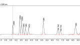
Given the above requirements for quality aggregate analysis, we selected a 1.8 µm SEC-3 column with a 300 Å nominal pore size and proprietary diol chemistry. The large pore size accommodates mAb, with a monomer size of approximately 5-6 nm, with dimers approach 9 nm [9]. Similar to the ion exchange based charge variant analysis, a bioinert column hardware can also minimise non-specific secondary adsorption in SEC and reduce the need for column priming. Therefore, the SEC column of choice also contains the bioinert titanium hardware. Using a mobile phase of 50 mM potassium phosphate and 250 mM potassium chloride at pH 6.8 with a flow rate 0.35 mL/min provided the data in Figure 4. Infliximab innovator provided >99% monomer, whereas biosimilar A and B contain 95% and 91% monomer, respectively.
Biosimilar development is growing in many markets globally and while regulatory guidelines give flexible parameters to demonstrate similarity, rigorous characterisation is still necessary. We’ve demonstrated two chromatographic techniques that can be employed to assess higher order structure of biosimilars relative to reference products. These analyses are enabled by judicious selection of appropriate particles for each technique as well as bioinert column hardware to minimise non-specific interactions.
1. US Food and Drug Administration Guidance for Industry: Scientific Consideration in Demonstrating Biosimilarity to a Reference Product; 2015. [accessed October 12, 2018]. https://www.fda.gov/downloads/drugs/guidances/ucm291128.pdf
2. European Medicines Agency, Committee for Medicinal Products for Human Use Guideline on similar biological medicinal products containing biotechnology-derived proteins as active substance: quality issues (revision 1) [Internet]. London, UK: European Medicines Agency; [Adopted on May 22, 2014; cited October 12, 2018] http://www.ema.europa.eu/docs/en_GB/document_library/Scientific_guideline/2014/06/WC500167838.pdf
3. Carson KL. Flexibility—the guiding principle for antibody manufacturing. Nat Biotech. 2005;23:1054–1058
4. Khawli LA, Goswami S, Hutchinson R, Kwong ZW, Yang J, Wang X, Yao Z, Sreedhara A, Cano T, Tesar D, et al. Charge variants in IgG1: Isolation, characterization, in vitro binding properties and pharmacokinetics in rats. MAbs. 2010;2:613–24.
5. He XZ, Que AH, Mo JJ. Analysis of charge heterogeneities in mAbs using imaged CE. Electropheresis. 2009;30:714–722
6. Fanali, S, Haddad, PR, Poole, C, Shoenmakers, P, Lloyd, DK. Liquid Chromatography: Fundamentals and Instrumentation, Elsevier, 2013
7. Arthur, KK, Gabrielson, JP, Kendrick, BS, Stoner, MR. Detection of Protein Aggregates by Sedimentation Velocity Analytical Ultracentrifugation (SV-AUC): Sources of Variability and Their Relative Importance. J. Pharm. Sci. 2009;98:3522-3539
8. Hong P, Koza S, Bouvier E S P, Size-exclusion chromatography for the analysis of protein biotherapeutics and their aggregates. J. Liq. Chromatogr. Relat. Technol. 2012;35:2923–2950
9. Lavoisier, A,Schlaeppi, JM. Early developability screen of therapeutic antibody candidates using Taylor dispersion analysis and UV area imaging detection. MAbs. 2014;7(1):77-83