Bioanalytical
Published over 5 years ago. See the latest and most current information on Bioanalytical.
Researchers are paying increasing attention to a critically important class of biomolecules: glycans. Although less studied than other major building blocks of life - proteins, lipids, and nucleic acids - complex and diverse glycans are essential to life and are omnipresent in organisms from archaea to humans [1]. Recent advances in glycoscience have shone new light on glycans and their role as key metabolic, structural, and physical components in biological structures. The potential of glycoengineering has been established, perhaps most significantly, in therapeutic antibody development.
This article outlines examples of the role glycans play clinically, the challenges of glycan analysis, summarises the tools available to elucidate their intricate structures, and presents data comparing traditional LC-MS workflows with the emerging technique of high-resolution ion mobility (HRIM) spectroscopy.
With knowledge of glycobiology expanding, the tremendous importance of the function of glycans in many branches of life science [2] including vital fields such as oncology [3], immunology [4], and infectious disease [5] is being elucidated. Moreover, developments in glycan analysis have coincided with an increased capacity to control glycosylation of specific biomolecules. Known as glycoengineering, this process involves manipulating glycosylation patterns, either by genetic modulation of specific glycosyltransferases or through chemical manipulation of glycoconjugates after biosynthesis [6].
Differences in glycosylation have been shown to affect therapeutic monoclonal antibody (mAb) activity in a number of ways [7,8]. As a key example, reduction or elimination of fucose monosaccharides from Fc domain glycans (also known as defucosylation) has been repeatedly shown to increase antibody-dependent cellular cytotoxicity (ADCC). Defucosylating antibody constructs leads to higher activation of natural killer (NK) as well as other immune cells. This, in turn, causes greater ADCC where the therapeutic mAb binds, including cancer cells [12,13]. Genentech (South San Francisco, California, USA) applied this strategy to develop Gazyva (obinutuzumab) to succeed its
$7 billion blockbuster CD20-targeting mAb, Rituxan (rituximab). Patients receiving Gazyva showed greater depth of remission and increased progression-free survival compared to those who received Rituxan [9].
Additionally, glycosylation variation in patients can correlate with treatment outcomes. In order to optimise treatments and clinical trial outcomes (e.g. patient stratification), it is critically important to have the ability to identify glycosylation profiles in a high throughput manner.
With respect to vaccine development, another example of the clinical impact of glycans includes the HIV-1 envelope proteins, which is richly decorated with glycan structures that help the virus evade recognition by the immune system. This so-called ‘glycan shield’ is an active area of research and serves as a potential target for vaccine development and broadly neutralising antibody production [10]. Similarly, the COVID-19 pandemic has brought attention to the role the SARS-CoV-2 spike protein glycosylation plays throughout viral infection and in therapeutic development [11,12]. Building on research into broadly neutralising antibodies against HIV [13], researchers are now also looking to apply a similar approach to coronaviruses [14].
To leverage the potential of glycoengineering and enhanced knowledge of glycosylation patterns, researchers are increasingly interested in utilising better tools and methods for quickly and accurately determining glycan structures. However, despite impressive advances, glycan analysis still remains challenging, largely due the nature of glycans themselves.
Arguably the principal structural challenge is that glycans are one of the most structurally diverse biomolecule families [2]. Though most mammalian glycans are built from around nine monosaccharide units, these monomers can be attached to one another through many different glycosidic linkages, including in a branched fashion [15]. Furthermore, glycans may range from one monosaccharide to many. Collectively, the number of possible glycan structures quickly expands with each additional monomer and branch. It is estimated that for a hexasaccharide there are, in total, ~1012 possible glycan structures, though they may not all occur in nature [16].
In addition, glycan assembly is not template-driven like DNA, RNA, and protein biosynthesis. Instead, glycans are made through interlaced networks of glycosyltransferases acting in the endoplasmic reticulum and Golgi. As a result, different glycan structures can occur at the same glycosylation position, generating biomolecules that are identical except for the glycans they carry (i.e. glycoforms). The distributions of glycoforms for a given glycoconjugate are also subject to change as glycosyltransferase expression changes.
Importantly, many of the monosaccharides that make up glycans are isomers of one another - glucose, galactose, and mannose being prime examples. This means that many different glycan structures can have the same mass, charge, and physical properties, but vary widely in function and recognition. Separation and identification of these complex isomeric materials can be exceedingly time-consuming and resource intensive.
As described above, glycan structures are inherently complex and heterogeneous. The go-to technique for glycan analysis has long been mass spectrometry (MS) - it offers the resolving power and sensitivity needed to elucidate specific glycan structures and glycoforms isolated from biological materials [17,18]. However, to achieve meaningful resolution between highly similar glycans within the same sample, separation techniques are needed ahead of MS analysis.
On a practical level, LC separation of highly similar biomolecules - like many glycans are - often requires extended run times, creating a potential bottleneck in analytical workflows. Even with long run times, some glycans behave so similarly that LC coelution cannot be avoided, which complicates or even prevents full structural assignment [19].
So, while LC-MS remains the standard workflow for glycan analysis, there is a pressing need for faster techniques with better resolving power.
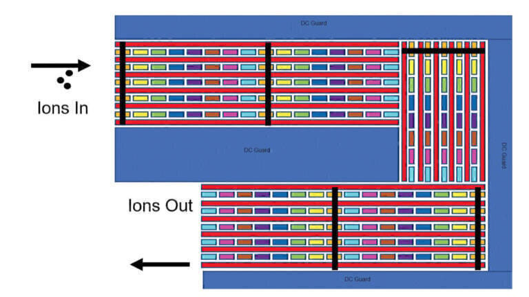
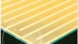
More recently, High Resolution Ion Mobility Mass Spectrometry (HRIM-MS) based on Structures for Lossless Ion Manipulation (SLIM) has emerged as a promising separation strategy in glycan analysis [20]. Unlike liquid chromatography, MOBILion’s HRIM system separates ionised molecules in the gas phase. The system uses printed circuit boards (PCBs), see Figure 1, held in a chamber maintained at constant pressure (2-4 torr). The PCBs have a series of radio frequency (RF), direct current (DC) and traveling wave electrodes printed on them that provide an ion conduit through which the analytes traverse along an exceptionally long serpentine path. The electric fields that propel the ions also prevent them from striking surfaces while moving, therefore preventing any losses along their way. Depending upon the speed of the traveling wave, ions either ‘surf’ and are not separated, or they undergo enough collisions with the gas molecules present that they roll over the traveling wave peaks, and separation occurs.
In HRIM, as with other ion mobility technologies, an ion’s migration time is determined by its mass-to-charge ratio and its size and shape. As ions are driven along the separation path; the collision with an inert buffer gas slows them down to a degree proportional to their size. The length of the ion separation path is crucial to achieving the high degree of separation required to gain adequate resolution of glycans with challenging structural diversity. MOBILion’s first HRIM product has an ion path of 13 meters (42 feet). The unique serpentine path design of the printed circuit boards allows for the 13m ion path to fit into a device the size of a briefcase allowing for unparalleled separation of isomeric structures.
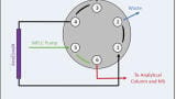
HRIM set-up is straightforward because it occurs in the gas phase under nearly ideal conditions. There is no need to optimise columns, flow rates, or liquid components as with LC. In addition, HRIM achieves rapid separations with data collection in the order of milliseconds to seconds. Much has been written about how the most advanced HRIM approaches boost resolving power by expanding the pathlength [21] and when combined with MS (HRIM-MS, Figure 2) offers tremendous potential for seamless separation of glycans followed by structural determination in biomedical and clinical research [22,23].
Recent work at the Complex Carbohydrates Research Center (CCRC) at the University of Georgia, Athens, Georgia, USA, has demonstrated the performance of HRIM-MS. Experiments compared this next-generation ion mobility technology, with an optimised LC-MS protocol [24].
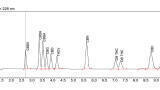
The experimental set-up at CCRC used the MOBILion HRIM instrument coupled to an Agilent 6545 Quadrupole Time of Flight (Q-ToF). Permethylated N-glycans released from Fetuin and RNaseB were analysed by 180-minute reverse phase nanoflow LC-MS/MS, typical of a traditional LC N-glycan analysis workflow. The same glycan species were analysed using HRIM-MS. In 2 minutes, all glycans identified using the 180-minute LC separation were separated and identified using the HRIM-MS workflow. This 90-fold time savings allows, for example, a three-month LC analysis to be compressed to a one-day analysis. Moreover, the HRIM-MS analysis resolved the structural isomers of a biantennary, disialylated complex glycan that could not be resolved with LC (Figure 3B, G5).
Being able to rapidly run hundreds of samples enables the establishment of a ‘normal range’ of glycan heterogeneity. That advantage allows greater insight into the behaviour of glycans in a wide range of illnesses, in a time frame compatible with the fast pace of biopharmaceutical development.
Glycan analysis, or glycomics, in which scientists elucidate the specific glycosylation patterns of biomolecules, has become an essential aspect of biomedical research, drug development, and biopharma quality control. Compared to traditional methods, HRIM excels in ease of use, generalisability of one method to multiple analyte classes, resolution, and speed of analysis. These attributes permit researchers to tackle the complexity of glycan structures and their heterogeneity on timescales that enable high-throughput analysis. The HRIM analyses discussed here are nearly 100-fold faster than LC analyses, but are only the beginning. More rapid sampling - a subject of ongoing development - could speed analysis times by additional orders of magnitude.
References
1. Varki A. Evolutionary forces shaping the Golgi glycosylation machinery: why cell surface glycans are universal to living cells. Cold Spring Harb Perspect Biol. 2011;3(6). doi: 10.1101/cshperspect.a005462.
PubMed PMID: 21525513; PMCID: PMC3098673.
2. Varki A. Biological roles of glycans. Glycobiology. 2017;27(1):3-49. doi: 10.1093/glycob/cww086. PubMed PMID: 27558841.
3. Stowell SR, Ju T, Cummings RD. Protein glycosylation in cancer. Ann Rev of Pathol-Mech, 2015;10(1):473–510. doi:10.1146/annurev-pathol-012414-040438
4. Baum LG, Cobb BA. The direct and indirect effects of glycans on immune function. Glycobiology. 2017;27(7):619–24.
5. Szymanski CM, Schnaar RL, Aebi M. Bacterial and viral infections. 2017. In: Varki A, Cummings RD, Esko JD, et al., editors. Essentials of Glycobiology [Internet]. 3rd edition. Cold Spring Harbor (NY): Cold Spring Harbor Laboratory Press; 2015-2017. Chapter 42. Available from: https://www.ncbi.nlm.nih.gov/books/NBK453060/ doi: 10.1101/glycobiology.3e.042
6. Kightlinger W, Warfel KF, DeLisa MP, Jewett MC. Synthetic Glycobiology: parts, systems, and applications. ACS Syn Bio. 2020;9(7):534-62.
7. Wang L-X, Tong X, Li C, Giddens JP, Li T. Glycoengineering of antibodies for modulating functions. Annu Rev Biochem. 2019; 88:26.1–26.27.
8. Jefferis R. Glycosylation as a strategy to improve antibody-based therapeutics. Nat Rev Drug Discov. 2009;8:226-34.
9. Ratner M. Genentech’s glyco-engineered antibody to succeed Rituxan. Nat Biotechnol. 2014;32(1):6-7.
10. Crispin M, Ward AB, Wilson IA. Structure and immune recognition of the HIV glycan shield. Annu Rev Biophys. 2018;47:499-523.
11. Watanabe Y, Allen JD, Wrapp D, McLellan JS, Crispin M. Site-specific glycan analysis of the SARSCoV- 2 spike. Science 2020;369:330-3.
12. Shajahan A, Supekar NT, Gleinich AS, Azadi P. Deducing the N- and O-glycosylation profile of the spike protein of novel coronavirus SARS-CoV-2. Glycobiology. 2020;1-8 doi: 10.1093/glycob/cwaa042
13. Shivatare VS, Shivatare SS, Lee CD, Liang CH, Liao KS, Cheng YY, Saidachary G, Wu CY, Lin NH, Kwong PD, Burton DR, Wu CY, Wong CH. Unprecedented role of hybrid N-glycans as ligands for HIV-1 broadly neutralizing antibodies. J Am Chem Soc. 2018;140(15):5202-10. doi: 10.1021/jacs.8b00896. PubMed PMID: 29578688.
14. Lv H, Wu NC, Tsang OT, Yuan M, Perera R, Leung WS, So RTY, Chan JMC, Yip GK, Chik TSH, Wang Y, Choi CYC, Lin Y, Ng WW, Zhao J, Poon LLM, Peiris JSM, Wilson IA, Mok CKP. Cross-reactive antibody response between SARS-CoV-2 and SARS-CoV Infections. Cell Rep. 2020;31(9):107725. Epub 2020/05/20. doi: 10.1016/j.celrep.2020.107725. PubMed PMID: 32426212; PMCID: PMC7231734.
15. Seeberger PH. Monosaccharide diversity. 2017. In: Varki A, Cummings RD, Esko JD, et al., editors. Essentials of Glycobiology [Internet]. 3rd edition. Cold Spring Harbor (NY): Cold Spring Harbor Laboratory Press; 2015-2017. Chapter 2. Available from: https://www.ncbi.nlm.nih.gov/books/NBK453086/ doi: 10.1101/glycobiology.3e.002
16. Laine RA. A calculation of all possible oligosaccharide isomers both branched and linear yields 1.05 x 10¹² structures for a reducing hexasaccharide: The Isomer Barrier to development of single-method saccharide sequencing or synthesis systems. Glycobiology. 1994;4(6):759-767.
doi:10.1093/glycob/4.6.759
17. Palaniappan KK, Bertozzi CR. Chemical Glycoproteomics. Chem Rev. 2016;116(23):14277-306. doi:10.1021/acs.chemrev.6b00023. PubMed PMID: 27960262; PMCID: PMC5327817.
18. Mulloy B, Dell A, Stanley P, et al. Structural analysis of glycans. 2017. In: Varki A, Cummings RD, Esko JD, et al., editors. Essentials of Glycobiology [Internet]. 3rd edition. Cold Spring Harbor (NY): Cold Spring Harbor Laboratory Press; 2015-2017. Chapter 50. Available from: https://www.ncbi.nlm.nih.gov/books/NBK453059/ doi: 10.1101/glycobiology.3e.050
19. Rudd P, Karlsson NG, Khoo KH, et al. Glycomics and Glycoproteomics. 2017. In: Varki A, Cummings RD, Esko JD, et al., editors. Essentials of Glycobiology [Internet]. 3rd edition. Cold Spring Harbor (NY): Cold Spring Harbor Laboratory Press; 2015-2017. Chapter 51. Available from: https://www.ncbi.nlm.nih.gov/books/NBK453015/ doi: 10.1101/glycobiology.3e.051
20. Hofmann J, Pagel K. Glycan Analysis by Ion Mobility-Mass Spectrometry. Angew Chem Int Edit. 2017;56(29):8342-9. doi: 10.1002/anie.201701309.
21. Hollerbach AL, Li A, Prabhakaran A, Nagy G, Harrilal CP, Conant CR, Norheim RV, Schimelfenig CE, Anderson GA, Garimella SVB, Smith RD, Ibrahim YM. Ultra-High-Resolution Ion Mobility Separations Over Extended Path Lengths and Mobility Ranges Achieved using a Multilevel Structures for Lossless Ion Manipulations Module. Anal Chem 2020;92(11):7972–9.
22. Arnaud CH. Resolving power for the people: Ion mobility-mass spec expands its offerings. C&EN. 2020;98(20).
23. Wormwood KL, Deng L, Hamid AM, DeBord D, Maxon L. The potential for Ion Mobility in pharmaceutical and clinical analyses. Advancements of Mass Spectrometry in Biomedical Research. 2019. p. 299-316.
24. Wormwood Moser KL, Arndt JR, Yadav A, Krufka S, Van Aken G, DeBord D, Webster G, Wells L, Tiemeyer M, Maxon L. Structures for Lossless Ion Manipulations (SLIM)-Mass Spectrometry (MS) for High Resolution and High Throughput Permethylated N-and O-Glycan Analysis. MOBILion Systems Inc. 2019.
Disclaimer: MOBILion is the exclusive licensee of the SLIM technology for commercialisation purposes. HRIM is intended for research use only. Not for diagnostic procedures.
For more information, visit MOBILion Systems Inc (https://mobilionsystems.com)
*Lance Wells is a Georgia Research Alliance Distinguished Investigator, Professor of Biochemistry and Molecular Biology, Director of integrated life sciences, and co-director of the Thermo Fisher appointed Center of Excellence in Glycoproteomics at the CCRC at the University of Georgia. He can be reached at [email protected].
** Michael Tiemeyer is a Distinguished Research Professor of Biochemistry and Molecular Biology, Co-director of the Thermo Fisher appointed Center of Excellence in Glycoproteomics at the University of Georgia, and Co-Director of the Complex Carbohydrate Research Center, University of Georgia. He can be reached at [email protected].
MOBILion Systems would also like to acknowledge Jeffrey Agar Ph.D., Associate Professor, Northeastern University, and Kelly L. Wormwood Moser Ph.D., Lab Operations Manager and Applications Scientist at MOBILion Systems Inc, for their input on this work.


-(1).jpg)