Bioanalytical
Published over 7 years ago. See the latest and most current information on Bioanalytical.
Many biopharmaceutical companies are working to develop next-generation biotherapeutics for the treatment of many, currently untreatable, diseases. In parallel, analytical methodologies have been developed to characterise such complex molecules. This article will discuss a new C4 superficially porous particle-packed column that has been optimised for characterising proteins, especially monoclonal antibodies and antibody-drug conjugates. Fundamental studies, looking at the chromatographic performance of the column, and application examples employing the column for biomolecule characterisation, will be highlighted.
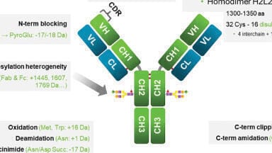
Monoclonal antibodies (mAbs) are manufactured by many biopharmaceutical companies to treat diseases ranging from Alzheimer’s disease, Parkinson’s disease, ulcerative colitis, and many types of cancers [1-3]. Most recombinant therapeutic mAbs belong to the human immunoglobulin G (IgG) category among the immunoglobulin superfamily. A schematic of an IgG antibody is depicted in Figure 1.
An IgG antibody is composed of two light chains (LC) that are tethered to two heavy chains (HC) through disulphide bonds. In addition, since the LC and HC are composed of amino acids with reactive side chains, IgG’s can be post-translationally modified through phosphorylation, methylation, oxidation, and nitrosylation, among other modifications. These modifications may change the binding affinity of the mAb so that it binds either the wrong antigen, does not bind any antigen, or associates with the wrong cell surface receptor. In addition, mAbs can also aggregate which can lead to allergic responses in patients. Biopharmaceutical companies need to develop rigorous methods to assess lot-to-lot reproducibility of their candidate biologic drug, and the above-mentioned modifications are known as Critical Quality Attributes (CQAs) that both the Food and Drug Administration (FDA) and the European Medicines Agency (EMA) monitor. Due to these stringent requirements from regulatory bodies, much research has been pursued in the past 20 years to develop accurate, robust, and high-throughput methods to assess biopharmaceutical purity and structure.
Ultra-high-performance liquid chromatography-mass spectrometry (UHPLC-MS) has emerged as a powerful technique to characterise these biomacromolecules. Most biopharmaceutical R&D laboratories, as well as quality control (QC) laboratories, have ready access to this type of instrumentation. Due to the lower system dispersion, lower dead volume, and higher upper pressure limit of these instruments, biopharmaceutical companies have been able to develop methods that not only probe the finest structural details of a candidate drug, but have enabled QC labs to assay hundreds of samples in a single day.
Besides advances in UHPLC and MS instrumentation, there have been many advances in the field of HPLC column and stationary phase development. Two main types of particle morphology are prevalent in the industry today: fully porous particles (FPPs) and superficially porous particles (SPPs, also called core-shell particles). To take advantage of the low dispersion of UHPLC instrumentation, columns with sub-2 μm FPPs with pore sizes of 300 Å have been used for the analysis of larger hydrodynamic radii biomacromolecules. These columns have been the industry standard since the mid 2000’s. However, these columns suffer limitations when analysing larger or more complex proteins like mAbs and antibody-drug conjugates (ADCs). The relatively small pore size, in addition to a totally porous architecture, and overall higher surface area, restricts the free diffusion of large molecules through the particle and may cause irreversible adsorption of the protein to the stationary phase. This architecture concomitantly results in an increase in the mass transfer term and longitudinal diffusion term of the van Deemter equation, leading to peak tailing, loss of resolution, and low recovery.
In recent years, the use of columns packed with SPPs has increased substantially. Historically, Horvath and Kirkland pioneered the concept and initial synthetic techniques for producing SPPs in the late 1960’s to early 1970’s [5,6]. The past 10 years have seen tremendous growth of these particles, offered by a range of manufacturers, and available for several different application areas [7]. These applications include small molecule pharmaceutical separations, pesticide analysis, glycan analysis, chiral separations, and large molecule separations.
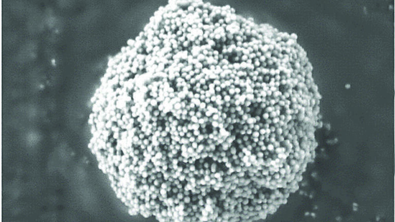
Current SPP column technology provide an alternative to sub-2 µm FPPs for biomolecule separations. Recently, a new 1000 Å C4 column has been introduced that has been optimised for mAb and ADC characterisation. This column is packed with 2.7 μm SPPs that are composed of a 0.5 μm shell thickness and a 1.7 μm solid silica core. The 1000 Å pore particle permits the analysis of mAbs, ADCs, and other, much larger, biomacromolecules. Figure 2 shows a scanning electron microscopy (SEM) image of an SPP. Note the presence of the solid silica core in the SPP. Advantages over columns packed with FPPs are numerous: the SPP shows a significant advantage in mass transfer, leading to less band spreading; columns packed with SPPs are more uniformly packed than columns composed of FPPs, leading to a lower eddy dispersion (A term) in the van Deemter equation; and larger particle sized SPPs have efficiencies similar to or better than sub-2 μm FPPs, leading to the ability of the analyst to run at higher flow rates with less risk of on-column frictional heating due to elevated column backpressure. Finally, the B-term (longitudinal diffusion) of the van Deemter equation is also minimised with SPPs. This is due to the presence of less dead volume in the column. A column packed with FPP particles will occupy only one-third of the column volume whereas a column packed with SPP particles will occupy approximately 25% more volume [8]. The major disadvantage of SPP particles is the lower loading capacity of these particles, which is a direct result of the lower overall pore volume. This characteristic of SPP particles leads to overloaded peaks if sample concentration and injection volume are not carefully considered. This same trait is why non-porous materials should also not be used for reversed-phase separations of proteins.
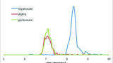
The gain in chromatographic performance observed when comparing SPP and FPP particle architecture, at elevated flow rate, is due mostly to the short diffusion path within the SPP architecture, thus enhancing mass transfer, in addition to less eddy diffusion. In addition, the BIOshellTM IgG column, as utilised here, has an average pore diameter of 1000 Å, thereby minimising any secondary size-exclusion effects that could lead to band broadening and loss of resolution. These concepts are illustrated in Figure 3 in which the SigmaMAb monoclonal antibody standard is assayed on three columns: BIOshellTM IgG 1000 Å C4, BIOshellTM A400 Protein C4, and a 1.7 μm, 300 Å FPP packed column. The BIOshellTM A400 Protein C4 column is a column packed with 3.4 μm, 400 Å, C4-bonded, SPP particles.
To ensure that this trend observed in Figure 3 was translatable to any mAb, a series of mAbs was assayed on the same columns, using the same chromatographic method, and the peak width at half height, a measure of chromatographic performance, was compared. Figure 4 summarises those results. None of the mAbs assayed generated peak widths, at half height, greater than 0.23 min on the BIOshellTM IgG 1000 Å C4 column whereas this value was consistently higher on the other two columns. It should be noted, as a caveat, that the particle sizes of the two BIOshellTM columns are different: the IgG and A400 columns have particles sizes of 2.7 and 3.4 μm, respectively. In addition, the shell thicknesses of the two particles are different: the IgG and A400 particles have shell thicknesses of 0.5 and 0.2 μm, respectively. A combination of both particle characteristics may be influencing why both BIOshellTM columns behave similarly. Also, the higher surface area of the FPP particles may be influencing why the FPP-packed column yielded higher peak-widths than the SPP-packed columns. The proteins may be partially or completely denatured due to interactions with the particle surface, and, due to different energetics between intact and denatured proteins, may be preferentially adsorbed to the stationary phase.
One method to gauge the efficiency of a column operating at high flow rates is to examine the peak volume of an analyte at varying flow rates. For SPPs, the mass transfer term of the van Deemter equation is relatively unaffected by flow rate. Thus, theoretically, peak volume should show relatively little change with increasing flow rate but should change (i.e. increase) for analytes assayed with FPP-packed columns. Using SigmaMAb as the analytical probe, this investigation was conducted using the BIOshell IgG 1000 Å C4 column, BIOshell A400 Protein C4, and the FPP, C4 columns used previously. Figure 5 summarises the results. The two BIOshell columns, as expected, show little change in peak volume with increasing flow rate while the FPP 300 column shows a steep increase due to the greater non-equilibrium effects caused by the increased diffusion paths associated with FPP architecture.
In antibody dependent cellular cytotoxicity (ADCC), FcγRs on the surface of effector cells (natural killer cells, macrophages, monocytes, and eosinophils) bind to the Fc region of an IgG which itself is bound to a target cell. Upon binding, a signalling pathway is triggered which results in the secretion of various substances, such as lytic enzymes, perforin, granzymes, and tumour necrosis factor, which mediate in the destruction of the target cell. The level of ADCC effector function varies for human IgG subtypes. Although this is dependent on the allotype and specific FcγR in simple terms, ADCC effector function is high for human IgG1 and IgG3, and low for IgG2 and IgG4 [9]. Because of their reduced effector function activity, IgG2 antibodies are becoming the favoured format for some protein therapeutics. All IgG2 biologics, however, are composed of different ratios of IgG2 isoforms that differ by the pattern of disulphide bonding in the hinge region. Figure 6 displays some of the possible isoforms of an IgG2 antibody [10].
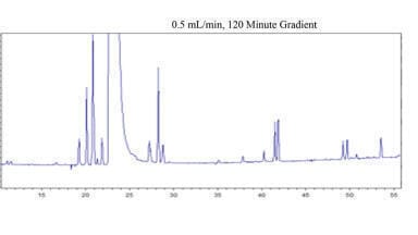
Because some of these isoforms may have immunogenic effects on a patient, and to ensure lot-to-lot reproducibility of a biologic, a method is required to resolve these different variants. Recently, an analytical reversed-phase chromatography (RPC) method was developed to resolve these different variants employing a BIOshellTM IgG 1000 Å C4 column. Figure 7 compares the chromatographic results of using a column packed with FPPs versus using the BIOshellTM IgG column in resolving different disulphide bond isoforms of Denosumab. Notice the improved resolution of disulphide bond isoforms using the BIOshellTM IgG column over the column packed with FPPs. Using this RPC method with the BIOshellTM IgG column, in combination with techniques such as redox amplification, thiol tagging, and mass spectrometry, it would be possible to identify and confirm the peaks in Figure 7.
Besides just using a mAb as the therapeutic, some companies are taking a mAb and tethering it to a cytotoxic drug, creating antibody-drug conjugates (ADCs). The modus operandi of these conjugate molecules is to use the site-specificity of an antibody to steer towards disease cells; once engulfed by a cell, the cytotoxic drug is released where it then elicits its therapeutic function.
However, the process of binding cytotoxic ‘payloads’ to an antibody creates a heterogeneous mixture of different permutations of a number of payloads tethered to the light or heavy chain of an antibody. Biopharmaceutical companies need to be able to identify how much payload is on the mAb, and where the payload is localised. The BIOshellTM IgG 1000 Å C4 column was employed to examine this. Figure 8 compares the results obtained on the 1000 Å column versus that with a SPP-polyphenyl phase chemistry. The higher peak capacity and efficiency realised with the 1000 Å C4 material over the polyphenyl material can clearly be observed. This application underscores the importance of not only pore size, but of stationary phase chemistry in eliciting enhanced separations of biomolecules. The method used in Figure 8 is mass spectrometry (MS) compatible, so the identity of each of the peaks can be deduced.
As the market for new drugs is slowly overtaken by biologics, the challenges in determining the purity of a new drug are many. New column technology, such as wide-pore SPP columns, will help scientists develop new methodologies to resolve these challenges. By reducing eddy dispersion and improving mass transfer, these columns yield tremendous gains in chromatographic performance compared with current FPP columns. As regulatory agencies require biopharmaceutical companies to add more CQAs in the monitoring of new drugs, this type of column technology will be useful.
1. H. M. Shepard, G. L. Philips, C. D. Thanos, and M. Feldmann, Clin. Med. (Lond.). 2017; 17: 220 – 232.
2. A. Hey, Microbiol. Spectr. 2015; 3, AID – 0026 – 2014.
3. J. C. Novak, A. E. Lovett-Racke, and M. K. Racke, Arch. Neurol. 2008; 65: 1162 – 1165.
4. C. E. Muraco, LC/GC North America, 35, 734-745 (2017)
5. C. G. Horvath, B. A. Preiss, S. R. Lipsky, Anal. Chem., 39, 1422-1428 (1967)
6. J. J. Kirkland, Anal. Chem. 41, 218-220 (1969)
7. J. J. Kirkland, F. A. Truszkowski, C. H. Dilks, and G. S. Engel, J. Chromatogr. A, 890 (2000), 3 – 13.
8. R. Hayes, A. Ahmed, T. Edge, and H. Zhang, J. Chromatogr. A, 1357 (2014), 36 – 52.
9. M. S. Powell and P. M. Hogarth, Multichain Immune Recognition Receptor Signaling. A. B. Sigalov, ed. (Springer New York), 22 – 34.
10. L. M. Jones, W. Cui, H. Zhang, M. L. Gross, J. Am. Soc. Mass Spectrom. 24, 835-842 (2013)