Autosamplers
Published over 11 years ago. See the latest and most current information on Autosamplers.
The post-translational modification of proteins with oligosaccharides, known as glycosylation, can affect both the structure and function of a protein [1-3]. Not surprisingly, the glycan profile of a biopharmaceutical is commonly defined as a critical quality attribute [4-5], since it can be a measure of efficacy and immunogenicity, as well as an indicator of manufacturing conditions [6-7]. N-glycosylation of proteins, such as monoclonal antibodies (mAbs), is consequently monitored during the development and lot release of biotherapeutics. Very often these assays are performed on N-glycans after they are released from their counterpart proteins [8]. Since glycans are highly polar molecular structures, they are amenable to hydrophilic interaction liquid chromatography (HILIC) based techniques for purification and separation (Figure 1). Rudd and co-workers have clearly shown the utility of HILIC for mapping the N-glycan heterogeneity of protein samples [9-15]. In such a method, the released N-glycans may be derivatised so that they bear a chromophore or fluorophore and can be detected optically following the chromatographic separation. Unfortunately, conventional approaches to the preparation of N-glycans for HILIC analysis are either laborious or require compromises in MS or optical sensitivity [16-17]. This limits the throughput or detail by which glycosylation can be characterised or monitored. In addition, many of these methods, due to their complexity, are challenging to transfer throughout an organisation. Thus, it would be advantageous for an N-glycan sample preparation protocol to be streamlined and robust, while facilitating sensitive MS and optical detection.
Accelerating Derivatisation
N-Glycan analysis methods often rely on an analyte derivatisation procedure that employs reductive amination. Reductive amination labels the reducing, aldehyde termini that form on N-glycans only after they hydrolyse from their glycosylamine form. This reaction requires an anhydrous reaction, which begins with a dry down step followed by a 2-3 hour chemical conversion process (Figure 2) [16-17].
These methods often contain numerous steps that require significant levels of expertise to optimise and because of the complex and stringent nature of this chemistry there is a high potential for analyst error which can result in repeating the sample preparation. To address this challenge, rapid tagging reagents have recently been introduced that can be used in place of conventional reductive amination labels, such as 2-aminobenzamide (2AB) [18]. These reagents target the native glycosylamines which are initially formed during enzymatic release from protein, to yield highly stable urea-linked derivatives. Although these reagents have accelerated labelling and quickly introduce a fluorophore for chromatographic detection, they have lacked the chemical properties needed to facilitate mass spectral identification of low abundance N-glycans. An improvement upon this approach for labelling released N-glycans would be designed to also derivatise the glycosylamine with a label that would allow for enhanced fluorescence and MS detectability. Figure 3 shows a purposefully designed compound called RapiFluor-MS™ that has these features.
In a 5 minute reaction, N-glycosylamines are labelled with RapiFluor-MS, a reagent comprised of an N-hydroxysuccinimide (NHS) carbamate rapid tagging group, a high quantum yield quinolone fluorophore, and a basic tertiary amine that enhances ionisation efficiency. This approach is dependent on a quick deglycosylation reaction that limits glycosylamine hydrolysis, a reaction that has been determined to have a half-life of approximately 2 hours at 50°C in the pH 7.9 digest buffer used. To this end, a rapid, PNGase F deglycosylation produces complete deglycosylation of most glycoproteins in 5 minutes. Coupled with nearly quantitative labelling it is predicted that less than 3% of the released glycosylamine will be unlabelled under the proposed conditions [19].
Building Robustness into N-Glycan Sample Preparations
Gains in efficiency and sensitivity are important, but so is having a protocol that is robust and able to produce consistent results. Every step in a protocol can potentially introduce bias and sample loss, for example a step to deplete the protein from the sample after deglycosylation. Not performing a protein removal step results in higher and more consistent glycan recovery when a molar excess of the labelling reagent based on glycoprotein concentration is employed. Studies have been performed with RapiFluor-MS labelling to show that the use of both higher and lower than recommended reagent produce comparable fluorescent profiles, indicating a high degree of robustness in the labelling (Figure 4).
While a clean-up step prior to labelling is avoided in this N-glycan sample preparation method, it is advantageous to have a clean-up step for the labelled glycans prior to HILIC chromatographic analysis, so that baseline interferences are minimised, resulting in improved relative quantitation from fluorescence chromatograms. However, extraction of the labelled glycans from reaction by-products is often a step that introduces bias. As mentioned previously, HILIC is an ideal mode for retaining glycans due to their highly polar nature. Therefore, a procedure has been optimised using a HILIC SPE process, involving a silica based aminopropyl sorbent. This particular stationary phase retains polar compounds with a surface that is both strongly hydrophilic and weakly basic, which allows an analyst to take advantage of electrostatic repulsion and ion exchange to improve yield and purity of the labelled N-glycans. The charge of the stationary phase surface is dependent on pH, at higher pH values the ionisation of basic analytes and the aminopropyl ligand is reduced, however the ionisation of the surface silanols is increased. Therefore, it is critical to choose an eluent that will have a fixed stable pH each time a sample preparation is performed (Figure 5).
Combining HILIC Chromatography with Mass Detection
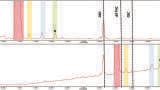
Finally, as with some traditional labelling chemistries such as 2-AB and 2-AA, RapiFluor-MS labelled glycans are ideally suited for HILIC chromatographic separations. UHPLC separations with a sub-2-µm amide bonded stationary phase such as the 2.1 x 150 mm ACQUITY UPLC BEH Amide 130Å 1.7µm provide the highest sample throughput. Figure 6 shows UHPLC chromatograms obtained for two samples prepared by different sample preparation techniques, one with 2-AB labelling and the other with RapiFluor-MS labelling. Notice that each set of labelled N-glycans are resolved by the HILIC separation with comparable selectivities. In this example, the mass load of RapiFluor-MS labelled glycans is lower given the enhanced sensitivity afforded by the label.
Due to the greatly improved MS sensitivity afforded by RapiFluor-MS, HILIC separation of labelled N-glycan samples can now be routinely analysed using LC combined with fluorescence (FLR) and MS detection. This capability provides high quality data for structural elucidation during characterisation, particularly for low abundance species. In addition, this dramatically improved MS sensitivity enables the opportunity to generate mass data during routine analyses often used in development, production, and quality control environments. Using conventional labelling technologies, the generation of meaningful mass data was only possible with high end mass spectrometers, since traditional reductive amination labels have inadequate ionisation efficiencies to facilitate alternative MS detection. With the introduction of RapiFluor-MS, which yields improved fluorescence sensitivity and dramatically improved MS sensitivity and correspondingly higher charge states in positive ion mode ESI-MS [19], it is now possible to use a simpler, lower cost mass detector for glycan MS analysis. An example of use of this workflow to characterise and routinely interrogate samples is shown in Figure 7. In this example, we see that a wide range of N-glycan structures are readily detected with both fluorescence and mass detectors. To further highlight the utility of mass detection, particularly in production environments, separation times can be reduced significantly while incorporating the use of selected ion recording, an intrinsic capability of the mass detector. In this way, critical species can be readily monitored without the need for extensive chromatographic separation. As shown in Figure 8, both MAN5 and FA2G1species can be independently monitored despite the fact that they co-elute in this analysis. The total analysis time is less than 10 minutes, and coupled with rapid labelling, results can be obtained from starting with the intact glycoprotein in less than 40 minutes.
Conclusion
By combining RapiFluor-MS labelling with rapid deglycosylation and a robust HILIC SPE micro-elution clean-up, an analyst can now complete a reproducible N-glycan sample preparation, from glycoprotein to ready-to-analyse sample, in just 30 minutes. Previous rapid and simple N-glycan sample preparation methods, which took approximately 3 hours to complete, required significant compromises to MS sensitivity. RapiFluor-MS labelling removes this limitation [19], thereby making it possible to routinely use mass detection to expedite accurate mass and MS-MS peak characterisation, or provide greater confidence in routine analyses using a mass detector.
References
1. Ohtsubo, K.; Marth, J. D., Cell 2006, 126 (5), 855-67.
2. Mechref, Y.; Hu, Y.; Garcia, A.; Zhou, S.; Desantos-Garcia, J. L.; Hussein, A., Bioanalysis 2012, 4 (20), 2457-69.
3. Ruhaak, L. R.; Miyamoto, S.; Lebrilla, C. B., Mol Cell Proteomics 2013, 12 (4), 846-55.
4. Glycosylation Engineering of Biopharmaceuticals. Humana Press: 2013.
5. Rathore, A. S.; Winkle, H., Nat Biotechnol 2009, 27 (1), 26-34.
6. Beck, A.; Wagner-Rousset, E.; Ayoub, D.; Van Dorsselaer, A.; Sanglier-Cianferani, S., Anal Chem 2013, 85 (2), 715-36.
7. Dalziel, M.; Crispin, M.; Scanlan, C. N.; Zitzmann, N.; Dwek, R. A., Science 2014, 343 (6166), 1235681.
8. Tarentino, A. L.; Gomez, C. M.; Plummer, T. H., Jr., Biochemistry 1985, 24 (17), 4665-71.
9. Rudd, P. M.; Guile, G. R.; Kuster, B.; Harvey, D. J.; Opdenakker, G.; Dwek, R. A., Nature 1997, 388 (6638), 205-7.
10. Tharmalingam, T.; Adamczyk, B.; Doherty, M. A.; Royle, L.; Rudd, P. M., Glycoconj J 2013, 30 (2), 137-46.
11. Houel, S.; Hilliard, M.; Yu, Y. Q.; McLoughlin, N.; Martin, S. M.; Rudd, P. M.; Williams, J. P.; Chen, W., Anal Chem 2014, 86 (1), 576-84.
12. Kaneshiro, K.; Watanabe, M.; Terasawa, K.; Uchimura, H.; Fukuyama, Y.; Iwamoto, S.; Sato, T. A.; Shimizu, K.; Tsujimoto, G.; Tanaka, K., Anal Chem 2012, 84 (16), 7146-51.
13. Ahn, J.; Bones, J.; Yu, Y. Q.; Rudd, P. M.; Gilar, M., J Chromatogr B Analyt Technol Biomed Life Sci 2010, 878 (3-4), 403-8.
14. Marino, K.; Bones, J.; Kattla, J. J.; Rudd, P. M., Nat Chem Biol 2010, 6 (10),
713-23.
15. Leymarie, N.; Zaia, J., Anal Chem 2012, 84 (7), 3040-8.
16. Mechref, Y.; Hu, Y.; Desantos-Garcia, J. L.; Hussein, A.; Tang, H., Mol Cell Proteomics 2013, 12 (4), 874-84.
17. Ruhaak, L. R.; Zauner, G.; Huhn, C.; Bruggink, C.; Deelder, A. M.; Wuhrer, M., Anal Bioanal Chem 2010, 397 (8), 3457-81.
18. Cook, K. S.; Bullock, K.; Sullivan, T., Biologicals 2012, 40 (2), 109-17.
19. Lauber, M. A.; Yu, Y. Q.; Brousmiche, D. W.; Hua, Z.; Koza, S. M.; Magnelli, P.; Guthrie, E.; Taron, C. H.; Fountain, K. J., Anal Chem 2015, DOI: 10.1021/acs.analchem.5b00758.