Autosamplers
A novel approach to measurement of hydrodynamic radius for a standard protein using UV area imaging detection
Oct 21 2009
Author: Alexander J S Chapman and David M Goodall on behalf of Paraytec Ltd
This article describes a novel approach to measurement of hydrodynamic radius for a standard protein using UV area imaging detection. The method allows proteins to be used in their native form, without any labelling or denaturation. A plug of protein solution is injected into a fused silica capillary, driven through the capillary by application of pressure, and detected using UV area imaging as it passes windows at entrance to and exit from a loop in the capillary. The radius of the protein is determined by analysis of band broadening due to Taylor dispersion. The method is applicable over a wide concentration range and uses only nanolitres of sample.
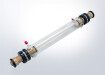
Overview of UV area imaging UV area imaging has been developed on a commercial basis by Paraytec Ltd through their patented ActiPixTM technology. The ActiPix is the world’s first UV area imaging detector, designed for on-line monitoring at a single UV wavelength, particularly for use with separation methods such as liquid chromatography (LC) and capillary electrophoresis (CE). The miniature size allows the detector to be used as a ‘plug and play’ accessory linked to existing separations instrumentation and in line with a mass spectrometer. Detection is performed at a selected wavelength by means of exchangeable filters. When light is shone
through a liquid-filled capillary, the liquid inside the capillary and the capillary vessel combine to act as a cylindrical lens. With a fused silica capillary, there is excellent light transmission in the UV down to 190 nm.
A key innovation of this technology is use of an area imaging array instead of the more usual linear photodiode array. This allows light passing through the sample in the capillary to be readily referenced against light from the same source. This process is illustrated using the ray diagram in Figure 1. The light focused through the centre of the capillary is represented in blue, the reference light is represented as yellow, and the boundary
between reference and sample is represented by the purple lines. The major benefit of using this self-referencing process is that it is independent of any light fluctuations, resulting in a very stable absorbance signal output.
Digital Edition
Chromatography Today - Buyers' Guide 2022
October 2023
In This Edition Modern & Practical Applications - Accelerating ADC Development with Mass Spectrometry - Implementing High-Resolution Ion Mobility into Peptide Mapping Workflows Chromatogr...
View all digital editions


Events
May 05 2024 Seville, Spain
May 15 2024 Birmingham, UK
May 19 2024 Brno, Czech Republic
May 21 2024 Lagos, Nigeria
May 23 2024 Beijing, China