Bioanalytical
Automation Arrives
Aug 25 2015
Author: ard Hopfgartner,n, Sandrine Cudby Emmanuel Varesio, Sandra Jahré, Gér on behalf of University of Geneva
Metabolomic studies provide us with key insights into biological systems and can help improve our understanding of the basis of a disease, elucidate the mechanism of action of a drug, and inform the development of relevant biomarkers. In practice, for metabolites to be studied, they need to be effectively extracted from a sample, a process that can be challenging due to their broad physiochemical diversities and the potentially large spectrum of concentrations of different metabolites in a single sample.
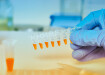
Liquid-liquid extraction (LLE) is often used to extract metabolites. However, the solvents employed as well as the experimental practice must be carefully considered in order to mitigate any risk affecting the quality of the resulting extract. Bligh and Dyer extraction, or one of its variants, such as Folch extraction, have been used for many years and is generally considered an efficient method for the extraction of lipids and polar endogenous metabolites from tissues and cells.1,2,3 However, it is still performed manually, and is a laborious task that is becoming increasingly out of place as laboratories move to automated analysis and handle large sets of samples.
In this article the authors describe recent work that details a fully automated Bligh and Dyer extraction and dual-column UHPLC-MS/MS separation for metabolomic analyses of tissues and cells, and compares the new procedure to a traditional manual method.
Advancing processes
Laboratories are constantly challenged to push the limits of analysis. New and improved measurement technology drives detection limits and time to deliver results down. As a consequence, bottlenecks and variability stemming from sample preparation have come under the spotlight. Automated approaches to sample prep are now routine but ‘smart sample preparation’ where robotics interfaces directly to an analyser, and software integration is also achieved, are much less common. In many laboratory settings automation has proven effective in increasing efficiency and improving repeatability. Streamlining workflows in this way not only increases consistency but also allows scientists to devote more time to operations that require their unique skills and experiences.
Full integration
Sample preparation workflows for metabolomic studies of tissues and cells often require a liquid-liquid extraction, which takes advantage of differing distribution coefficients to enrich metabolites and to separate them from undesired compounds. Now, an automated Bligh and Dyer extraction on a robotic system has been fully integrated with a dual-column ultra high-pressure liquid chromatography tandem mass spectrometer (UHPLC-MS/MS) platform for the metabolomics analysis of tissues and/or cells (Figure 1).
Bligh and Dyer
Offline methodology
Bligh and Dyer extraction involves a three step solvent extraction process, which traditionally must be completed by hand. The method can be adapted for complex lipid chemistries but typically the steps could include: 1) Treating the sample with methanol and chloroform, 2) chloroform only and then 3) with the addition of water. These steps ensure that any non-lipid contaminants also extracted are removed from the recovered lipids by washing or other solvent partition procedures (step 3) before the sample can undergo more detailed analysis. At each stage precautions have to be taken to minimize the risk of autoxidation of unsaturated fatty acids or hydrolysis of lipids, adding to the laboriousness of the method.
Instrumentation and software set-up
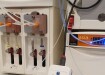
For the LC-separation of the Bligh and Dyer fractions, two quaternary low-pressure Nexera LC30AD UHPLC pumps (Shimadzu, Tokyo, Japan) were used. A PAL RTC robot (CTC Analytics, Zwingen, Switzerland) performed the sample prep (as outlined in figure 2), and injected the resulting aqueous fractions onto a 100 x 2.1 mm XBridge BEH 3.5 µm C18 XP column (Waters, Milford, MA, USA) running alternately with an acidic or a basic mobile phase. The organic (CHCl3) fractions were evaporated to dryness, reconstituted and injected onto a 150 x 2.1 mm XBridge BEH C8 XP column. Both columns were maintained at 40°C.
The dual-column UHPLC platform was hyphenated to a TripleTOF 5600 MS (AB Sciex, Ontario, Canada). Mass spectrometry and tandem mass spectrometry data were acquired in positive mode using a Turbo V ion source equipped with an APCI probe for automated calibration.
To complement the hardware configuration and allow for true integration the PAL RTC robot was controlled by PAL Sample Control v 2.1 software (CTC Analytics). This interface was linked with the LabSolutions v 5.6 SP2 software (Shimadzu) and used to control the UHPLC system.
Sample preparation
Figure 2 shows the sample preparation workflow for the offline and automated Bligh and Dyer extraction. The method proved to be far more streamlined than conventional sample preparation. Importantly, scientists only had to perform a few manual steps before loading samples onto the PAL RTC robot: initially 1 mL of cold MeOH was added to the raw samples, which were then spun ahead of flash freezing and cryogenic grinding. After the addition of a solution of H2O:MeOH:CHCl3 (0.8:2:1, v:v) the sample was then transferred to the PAL RTC robot where it underwent the automated Bligh and Dyer extraction.
The PAL RTC platform performed all the necessary extraction steps, splitting the fractions and adding the correct volumes and concentrations of the appropriate reagents and chromatographic standards as needed. Firstly, 225 μL H2O + 225 μL CHCl3 were added to the samples which were then spun in the centrifuge for 5 minutes at 4000 rpm.
500 µL of the upper fraction were then diluted 5-fold by adding 60 µL to 240 µL of dilution solvent (10% MeOH in samples). Chromatographic standards were then added and 25 µL injected on the C18 column (UHPLC system 1). Meanwhile 250 µL of the lower (CHCl3) fraction was evaporated to dryness with N2 gas (0.35 bar, 35°C for 10 minutes). The sample was then reconstituted in 150 µL MeOH. Again chromatographic standards were added and 5 µL was then injected on the C8 column (UHPLC system 2). The method proved significantly less labour intensive and reduced the overall sample preparation time from ca. 3 hours to prepare a dozen of samples to ca. half an hour for the offline preparation, since the other steps are prepared by the PAL RTC during the different LC runs without manual overhead time.
Analysis of the upper water-methanol fractions was conducted on UHPLC system 1 with on-line dilution and standard addition by the PAL RTC and alternating the pH of the mobile phases. The water-methanol Bligh and Dyer fractions contain 50% MeOH, thus an on-line dilution is necessary to lower the organic content in the sample solvent. Different dilution factors, with their adapted injection volumes to keep the same amount injected on-column, were tested with the PAL RTC. It was found that for polar compounds the peak widths increased for injections of more than 5 µL, as shown by the trace of adenine on Figure 3. However the peak area ratio for individual compounds remained adequate with up to 10% MeOH in the sample solvent, even with an injection volume of 45 µL. For lipophilic compounds peak widths remained at 0.1 min with large injections of 45 µL, but higher organic content in the sample solvent was required.
To account for the range in pKa-values (Table 1) the upper (aqueous) fractions were analysed with two different mobile phases (pH 3.0 and pH 8.3) to improve the retention of certain polar compounds, such as adenine and nicotine (Figure 3). When the mobile phases were alternated, the column reconditioning time for the basic pH was critical to ensure the stability of retention times. Therefore injection scheme B was preferred as each injection was alternated, as shown in Figure 4. The use of two pH conditions should ensure the compound is in a unique state and so will avoid issues with assay stability due to the compound being too close to its pKa. For compounds that have multiple pKas this approach should be reviewed with consideration for the revised pH conditions to avoid shifting retemtion times.
For highly polar compounds, high organic content or a large injection volume was detrimental to their peak shape. Conversely, for lipophilic compounds, losses were observed due to poor solubility in mostly aqueous solvents, such as those including only 5 or 10% methanol. However, for other compounds, consistent results were observed across the dilution factors.
The lower organic fraction was analysed on UHPLC system 2. With the automated system, evaporation of 400 µL in 1.2 mL vase vials was achieved in less than seven minutes for CHCl3 and less than 14 minutes for MeOH. After evaporation reconstituted volumes of 100, 150 and 200 µL of MeOH were tested for the organic fraction by injecting 5 µL on the column. It was found that 150 µL were sufficiently concentrated so this sample was chosen for further testing.
The automated Bligh and Dyer extractions lead to efficient separation of polar from unpolar metabolites. Only few compounds with amphiphilic properties, such as metoprolol, were retrieved in both fractions. These results are in agreement with comparative analyses of manually extracted samples.
In order to assess the repeatability of the automated Bligh and Dyer extraction process with the PAL RTC platform manual procedures were performed, which also allowed for the comparison of any variation between the two methods. Multiple extractions (n=5) of Chlamydomonas reinhardtii algae were performed. From the analyses of aqueous and organic fractions 20 variables were selected randomly by means of Marker View and/or Peak View software (only monoisotopic peaks with S/N > 30 were considered) and coefficients of variation (CV) were calculated for the corresponding peak areas. It was found that lower variation of CV <22% and therefore better repeatability was achieved with the automated method throughout all experiments conducted, as shown in Figure 5. This result was especially pronounced for the LC-MS runs of aqueous fractions at pH 8 and of the organic fractions.
By acquiring LC-MS/MS data in SWATH mode, the results could be directly subjected to a library search without the need of performing further targeted experiments, allowing for the fast identification and quantification of certain unknowns. This significantly reduced experimental time and resulted in faster processing of results.
Digital Edition
Chromatography Today - Buyers' Guide 2022
October 2023
In This Edition Modern & Practical Applications - Accelerating ADC Development with Mass Spectrometry - Implementing High-Resolution Ion Mobility into Peptide Mapping Workflows Chromatogr...
View all digital editions


Events
Apr 23 2024 Kintex, South Korea
Apr 23 2024 Seoul, South Korea
Apr 28 2024 Montreal, Quebec, Canada
May 05 2024 Seville, Spain
May 15 2024 Birmingham, UK